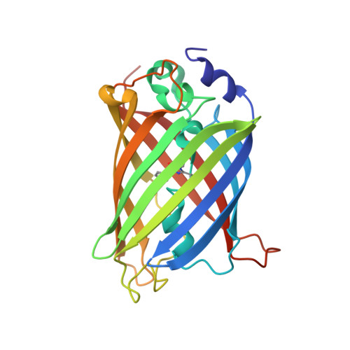
4L13

Crystal structure of Ligand Free EGFP-based Calcium Sensor CatchER


Experimental Data Snapshot

  • Method: X-RAY DIFFRACTION
  • Resolution: 1.66 Å
  • R-Value Free: 0.203 
  • R-Value Work: 0.181 
  • R-Value Observed: 0.182 

Starting Model: experimental
View more details

wwPDB Validation   3D Report Full Report


This is version 1.4 of the entry. See complete history


Literature

Structural basis for a hand-like site in the calcium sensor CatchER with fast kinetics.

Zhang, Y.Reddish, F.Tang, S.Zhuo, Y.Wang, Y.F.Yang, J.J.Weber, I.T.

(2013) Acta Crystallogr D Biol Crystallogr 69: 2309-2319

  • DOI: https://doi.org/10.1107/S0907444913021306
  • Primary Citation of Related Structures:  
    4L12, 4L13, 4L1I

  • PubMed Abstract: 

    Calcium ions, which are important signaling molecules, can be detected in the endoplasmic reticulum by an engineered mutant of green fluorescent protein (GFP) designated CatchER with a fast off-rate. High resolution (1.78-1.20 Å) crystal structures were analyzed for CatchER in the apo form and in complexes with calcium or gadolinium to probe the binding site for metal ions. While CatchER exhibits a 1:1 binding stoichiometry in solution, two positions were observed for each of the metal ions bound within the hand-like site formed by the carboxylate side chains of the mutated residues S147E, S202D, Q204E, F223E and T225E that may be responsible for its fast kinetic properties. Comparison of the structures of CatchER, wild-type GFP and enhanced GFP confirmed that different conformations of Thr203 and Glu222 are associated with the two forms of Tyr66 of the chromophore which are responsible for the absorbance wavelengths of the different proteins. Calcium binding to CatchER may shift the equilibrium for conformational population of the Glu222 side chain and lead to further changes in its optical properties.


  • Organizational Affiliation

    Department of Chemistry, Georgia State University, Atlanta, GA 30303, USA.


Macromolecules
Find similar proteins by:  (by identity cutoff)  |  3D Structure
Entity ID: 1
MoleculeChains Sequence LengthOrganismDetailsImage
EGFP-based Calcium Sensor CatchER230Aequorea victoriaMutation(s): 9 
Gene Names: GFP
UniProt
Find proteins for P42212 (Aequorea victoria)
Explore P42212 
Go to UniProtKB:  P42212
Entity Groups  
Sequence Clusters30% Identity50% Identity70% Identity90% Identity95% Identity100% Identity
UniProt GroupP42212
Sequence Annotations
Expand
  • Reference Sequence
Small Molecules
Ligands 1 Unique
IDChains Name / Formula / InChI Key2D Diagram3D Interactions
ACY
Query on ACY

Download Ideal Coordinates CCD File 
B [auth A]ACETIC ACID
C2 H4 O2
QTBSBXVTEAMEQO-UHFFFAOYSA-N
Modified Residues  1 Unique
IDChains TypeFormula2D DiagramParent
CRO
Query on CRO
A
L-PEPTIDE LINKINGC15 H17 N3 O5THR, TYR, GLY
Experimental Data & Validation

Experimental Data

  • Method: X-RAY DIFFRACTION
  • Resolution: 1.66 Å
  • R-Value Free: 0.203 
  • R-Value Work: 0.181 
  • R-Value Observed: 0.182 
  • Space Group: C 2 2 21
Unit Cell:
Length ( Å )Angle ( ˚ )
a = 61.401α = 90
b = 88.534β = 90
c = 118.427γ = 90
Software Package:
Software NamePurpose
SERGUIdata collection
MOLREPphasing
REFMACrefinement
HKL-2000data reduction
HKL-2000data scaling

Structure Validation

View Full Validation Report


Entry History 

Deposition Data

Revision History  (Full details and data files)

  • Version 1.0: 2013-12-11
    Type: Initial release
  • Version 1.1: 2014-01-08
    Changes: Database references
  • Version 1.2: 2023-09-20
    Changes: Data collection, Database references, Derived calculations, Refinement description
  • Version 1.3: 2023-12-06
    Changes: Data collection
  • Version 1.4: 2024-11-20
    Changes: Structure summary