4RER

Crystal structure of the phosphorylated human alpha1 beta2 gamma1 holo-AMPK complex bound to AMP and cyclodextrin


Experimental Data Snapshot

  • Method: X-RAY DIFFRACTION
  • Resolution: 4.05 Å
  • R-Value Free: 0.260 
  • R-Value Work: 0.224 
  • R-Value Observed: 0.227 

Starting Model: experimental
View more details

wwPDB Validation   3D Report Full Report


This is version 2.2 of the entry. See complete history


Literature

Structural basis of AMPK regulation by adenine nucleotides and glycogen.

Li, X.Wang, L.Zhou, X.E.Ke, J.de Waal, P.W.Gu, X.Tan, M.H.Wang, D.Wu, D.Xu, H.E.Melcher, K.

(2015) Cell Res 25: 50-66

  • DOI: https://doi.org/10.1038/cr.2014.150
  • Primary Citation of Related Structures:  
    4RED, 4RER, 4REW

  • PubMed Abstract: 

    AMP-activated protein kinase (AMPK) is a central cellular energy sensor and regulator of energy homeostasis, and a promising drug target for the treatment of diabetes, obesity, and cancer. Here we present low-resolution crystal structures of the human α1β2γ1 holo-AMPK complex bound to its allosteric modulators AMP and the glycogen-mimic cyclodextrin, both in the phosphorylated (4.05 Å) and non-phosphorylated (4.60 Å) state. In addition, we have solved a 2.95 Å structure of the human kinase domain (KD) bound to the adjacent autoinhibitory domain (AID) and have performed extensive biochemical and mutational studies. Together, these studies illustrate an underlying mechanism of allosteric AMPK modulation by AMP and glycogen, whose binding changes the equilibria between alternate AID (AMP) and carbohydrate-binding module (glycogen) interactions.


  • Organizational Affiliation

    1] Key Laboratory of Regenerative Biology, Guangzhou Institute of Biomedicine and Health, Chinese Academy of Sciences, Guangzhou, Guangdong 510530, China [2] School of Life Science, University of Science and Technology of China, Hefei, Anhui 230027, China [3] Laboratory of Structural Sciences, Van Andel Research Institute, 333 Bostwick Ave, NE, Grand Rapids, MI 49503, USA.


Macromolecules
Find similar proteins by:  (by identity cutoff)  |  3D Structure
Entity ID: 1
MoleculeChains Sequence LengthOrganismDetailsImage
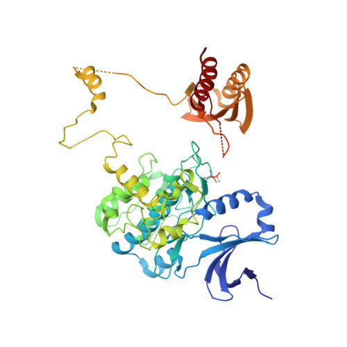
5'-AMP-activated protein kinase catalytic subunit alpha-1540Homo sapiensMutation(s): 3 
Gene Names: AMPK1Human holo-AMPK alpha1 subunitPRKAA1
EC: 2.7.11.1 (PDB Primary Data), 2.7.11.27 (PDB Primary Data), 2.7.11.31 (PDB Primary Data), 2.7.11.26 (PDB Primary Data)
UniProt & NIH Common Fund Data Resources
Find proteins for Q13131 (Homo sapiens)
Explore Q13131 
Go to UniProtKB:  Q13131
PHAROS:  Q13131
GTEx:  ENSG00000132356 
Entity Groups  
Sequence Clusters30% Identity50% Identity70% Identity90% Identity95% Identity100% Identity
UniProt GroupQ13131
Sequence Annotations
Expand
  • Reference Sequence
Find similar proteins by:  (by identity cutoff)  |  3D Structure
Entity ID: 2
MoleculeChains Sequence LengthOrganismDetailsImage
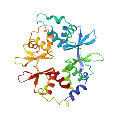
5'-AMP-activated protein kinase subunit beta-2197Homo sapiensMutation(s): 0 
Gene Names: Human holo-AMPK beta2 subunitPRKAB2
UniProt & NIH Common Fund Data Resources
Find proteins for O43741 (Homo sapiens)
Explore O43741 
Go to UniProtKB:  O43741
PHAROS:  O43741
GTEx:  ENSG00000131791 
Entity Groups  
Sequence Clusters30% Identity50% Identity70% Identity90% Identity95% Identity100% Identity
UniProt GroupO43741
Sequence Annotations
Expand
  • Reference Sequence
Find similar proteins by:  (by identity cutoff)  |  3D Structure
Entity ID: 3
MoleculeChains Sequence LengthOrganismDetailsImage
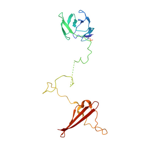
5'-AMP-activated protein kinase subunit gamma-1C [auth G]304Homo sapiensMutation(s): 0 
Gene Names: Human holo-AMPK gamma1 subunitPRKAG1
UniProt & NIH Common Fund Data Resources
Find proteins for P54619 (Homo sapiens)
Explore P54619 
Go to UniProtKB:  P54619
PHAROS:  P54619
GTEx:  ENSG00000181929 
Entity Groups  
Sequence Clusters30% Identity50% Identity70% Identity90% Identity95% Identity100% Identity
UniProt GroupP54619
Sequence Annotations
Expand
  • Reference Sequence
Oligosaccharides

Help

Entity ID: 4
MoleculeChains Length2D Diagram Glycosylation3D Interactions
Cycloheptakis-(1-4)-(alpha-D-glucopyranose)D [auth C]7N/A
Glycosylation Resources
GlyTouCan:  G01435GL
GlyCosmos:  G01435GL
Biologically Interesting Molecules (External Reference) 1 Unique
Experimental Data & Validation

Experimental Data

  • Method: X-RAY DIFFRACTION
  • Resolution: 4.05 Å
  • R-Value Free: 0.260 
  • R-Value Work: 0.224 
  • R-Value Observed: 0.227 
  • Space Group: P 32 2 1
Unit Cell:
Length ( Å )Angle ( ˚ )
a = 132.566α = 90
b = 132.566β = 90
c = 195.392γ = 120
Software Package:
Software NamePurpose
HKL-2000data collection
PHASERphasing
PHENIXrefinement
HKL-2000data reduction
HKL-2000data scaling

Structure Validation

View Full Validation Report


Entry History 

Deposition Data

Revision History  (Full details and data files)

  • Version 1.0: 2014-12-10
    Type: Initial release
  • Version 1.1: 2015-01-14
    Changes: Database references
  • Version 1.2: 2015-03-11
    Changes: Structure summary
  • Version 2.0: 2020-07-29
    Type: Remediation
    Reason: Carbohydrate remediation
    Changes: Atomic model, Data collection, Database references, Derived calculations, Non-polymer description, Structure summary
  • Version 2.1: 2023-09-20
    Changes: Data collection, Database references, Refinement description, Structure summary
  • Version 2.2: 2024-11-06
    Changes: Structure summary