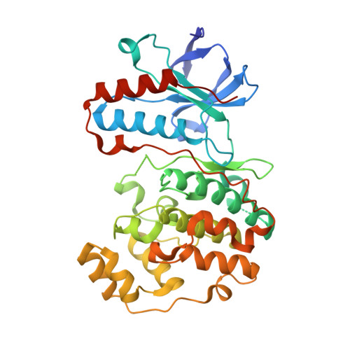
5R9Y

PanDDA analysis group deposition Form1 MAP kinase p38-alpha -- Fragment N13619a in complex with MAP kinase p38-alpha


Experimental Data Snapshot

  • Method: X-RAY DIFFRACTION
  • Resolution: 1.57 Å
  • R-Value Free: 0.210 
  • R-Value Work: 0.190 
  • R-Value Observed: 0.191 

wwPDB Validation   3D Report Full Report


Ligand Structure Quality Assessment 


This is version 1.2 of the entry. See complete history


Literature

Mining the PDB for Tractable Cases Where X-ray Crystallography Combined with Fragment Screens Can Be Used to Systematically Design Protein-Protein Inhibitors: Two Test Cases Illustrated by IL1 beta-IL1R and p38 alpha-TAB1 Complexes.

Nichols, C.Ng, J.Keshu, A.Kelly, G.Conte, M.R.Marber, M.S.Fraternali, F.De Nicola, G.F.

(2020) J Med Chem 63: 7559-7568

  • DOI: https://doi.org/10.1021/acs.jmedchem.0c00403
  • Primary Citation of Related Structures:  
    5R85, 5R86, 5R87, 5R88, 5R89, 5R8A, 5R8B, 5R8C, 5R8D, 5R8E, 5R8F, 5R8G, 5R8H, 5R8I, 5R8J, 5R8K, 5R8L, 5R8M, 5R8N, 5R8O, 5R8P, 5R8Q, 5R8U, 5R8V, 5R8W, 5R8X, 5R8Y, 5R8Z, 5R90, 5R91, 5R92, 5R93, 5R94, 5R95, 5R96, 5R97, 5R98, 5R99, 5R9A, 5R9B, 5R9C, 5R9D, 5R9E, 5R9F, 5R9G, 5R9H, 5R9I, 5R9J, 5R9K, 5R9L

  • PubMed Abstract: 

    Nowadays, it is possible to combine X-ray crystallography and fragment screening in a medium throughput fashion to chemically probe the surfaces used by proteins to interact and use the outcome of the screens to systematically design protein-protein inhibitors. To prove it, we first performed a bioinformatics analysis of the Protein Data Bank protein complexes, which revealed over 400 cases where the crystal lattice of the target in the free form is such that large portions of the interacting surfaces are free from lattice contacts and therefore accessible to fragments during soaks. Among the tractable complexes identified, we then performed single fragment crystal screens on two particular interesting cases: the Il1β-ILR and p38α-TAB1 complexes. The result of the screens showed that fragments tend to bind in clusters, highlighting the small-molecule hotspots on the surface of the target protein. In most of the cases, the hotspots overlapped with the binding sites of the interacting proteins.


  • Organizational Affiliation

    British Heart Foundation Centre of Excellence, Department of Cardiology, The Rayne Institute, St Thomas' Hospital, King's College London, London SE1 7EH, U.K.


Macromolecules
Find similar proteins by:  (by identity cutoff)  |  3D Structure
Entity ID: 1
MoleculeChains Sequence LengthOrganismDetailsImage
Mitogen-activated protein kinase 14360Mus musculusMutation(s): 0 
Gene Names: Mapk14Crk1Csbp1Csbp2
EC: 2.7.11.24
UniProt
Find proteins for P47811 (Mus musculus)
Explore P47811 
Go to UniProtKB:  P47811
Entity Groups  
Sequence Clusters30% Identity50% Identity70% Identity90% Identity95% Identity100% Identity
UniProt GroupP47811
Sequence Annotations
Expand
  • Reference Sequence
Small Molecules
Ligands 4 Unique
IDChains Name / Formula / InChI Key2D Diagram3D Interactions
SU7
Query on SU7

Download Ideal Coordinates CCD File 
B [auth A]3-oxidanylidene-3-[4-(phenylmethyl)piperidin-1-yl]propanenitrile
C15 H18 N2 O
ZCRMYTZFCMQXHR-UHFFFAOYSA-N
SO4
Query on SO4

Download Ideal Coordinates CCD File 
H [auth A]SULFATE ION
O4 S
QAOWNCQODCNURD-UHFFFAOYSA-L
DMS
Query on DMS

Download Ideal Coordinates CCD File 
I [auth A]DIMETHYL SULFOXIDE
C2 H6 O S
IAZDPXIOMUYVGZ-UHFFFAOYSA-N
CL
Query on CL

Download Ideal Coordinates CCD File 
C [auth A],
D [auth A],
E [auth A],
F [auth A],
G [auth A]
CHLORIDE ION
Cl
VEXZGXHMUGYJMC-UHFFFAOYSA-M
Experimental Data & Validation

Experimental Data

  • Method: X-RAY DIFFRACTION
  • Resolution: 1.57 Å
  • R-Value Free: 0.210 
  • R-Value Work: 0.190 
  • R-Value Observed: 0.191 
  • Space Group: P 21 21 21
Unit Cell:
Length ( Å )Angle ( ˚ )
a = 45.72α = 90
b = 87.21β = 90
c = 125.65γ = 90
Software Package:
Software NamePurpose
REFMACrefinement
Aimlessdata scaling
PDB_EXTRACTdata extraction
XDSdata reduction
REFMACphasing

Structure Validation

View Full Validation Report


Ligand Structure Quality Assessment 


Entry History 

Deposition Data

Revision History  (Full details and data files)

  • Version 1.0: 2020-07-22
    Type: Initial release
  • Version 1.1: 2020-08-05
    Changes: Database references
  • Version 1.2: 2024-03-06
    Changes: Data collection, Database references