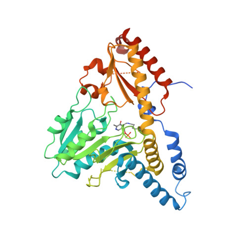
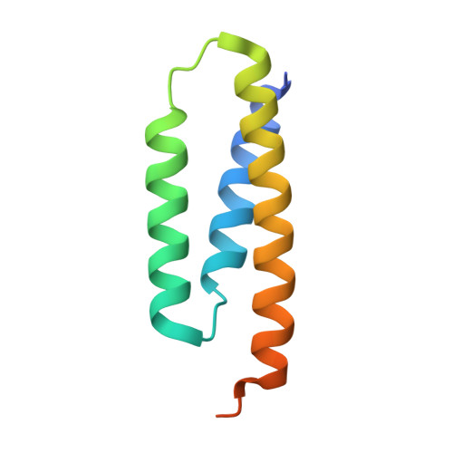
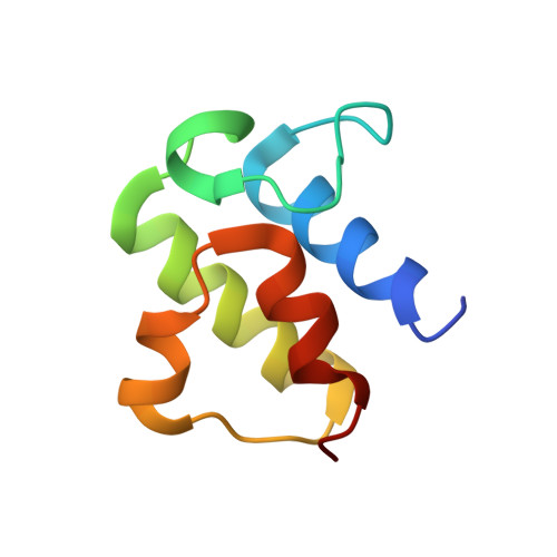
5USR

Crystal structure of human NFS1-ISD11 in complex with E. coli acyl-carrier protein at 3.09 angstroms


Experimental Data Snapshot

  • Method: X-RAY DIFFRACTION
  • Resolution: 3.09 Å
  • R-Value Free: 0.259 
  • R-Value Work: 0.212 
  • R-Value Observed: 0.214 

Starting Model: experimental
View more details

wwPDB Validation   3D Report Full Report


Ligand Structure Quality Assessment 


This is version 1.6 of the entry. See complete history


Literature

Structure of human Fe-S assembly subcomplex reveals unexpected cysteine desulfurase architecture and acyl-ACP-ISD11 interactions.

Cory, S.A.Van Vranken, J.G.Brignole, E.J.Patra, S.Winge, D.R.Drennan, C.L.Rutter, J.Barondeau, D.P.

(2017) Proc Natl Acad Sci U S A 114: E5325-E5334

  • DOI: https://doi.org/10.1073/pnas.1702849114
  • Primary Citation of Related Structures:  
    5USR

  • PubMed Abstract: 

    In eukaryotes, sulfur is mobilized for incorporation into multiple biosynthetic pathways by a cysteine desulfurase complex that consists of a catalytic subunit (NFS1), LYR protein (ISD11), and acyl carrier protein (ACP). This NFS1-ISD11-ACP (SDA) complex forms the core of the iron-sulfur (Fe-S) assembly complex and associates with assembly proteins ISCU2, frataxin (FXN), and ferredoxin to synthesize Fe-S clusters. Here we present crystallographic and electron microscopic structures of the SDA complex coupled to enzyme kinetic and cell-based studies to provide structure-function properties of a mitochondrial cysteine desulfurase. Unlike prokaryotic cysteine desulfurases, the SDA structure adopts an unexpected architecture in which a pair of ISD11 subunits form the dimeric core of the SDA complex, which clarifies the critical role of ISD11 in eukaryotic assemblies. The different quaternary structure results in an incompletely formed substrate channel and solvent-exposed pyridoxal 5'-phosphate cofactor and provides a rationale for the allosteric activator function of FXN in eukaryotic systems. The structure also reveals the 4'-phosphopantetheine-conjugated acyl-group of ACP occupies the hydrophobic core of ISD11, explaining the basis of ACP stabilization. The unexpected architecture for the SDA complex provides a framework for understanding interactions with acceptor proteins for sulfur-containing biosynthetic pathways, elucidating mechanistic details of eukaryotic Fe-S cluster biosynthesis, and clarifying how defects in Fe-S cluster assembly lead to diseases such as Friedreich's ataxia. Moreover, our results support a lock-and-key model in which LYR proteins associate with acyl-ACP as a mechanism for fatty acid biosynthesis to coordinate the expression, Fe-S cofactor maturation, and activity of the respiratory complexes.


  • Organizational Affiliation

    Department of Chemistry, Texas A&M University, College Station, TX 77842.


Macromolecules
Find similar proteins by:  (by identity cutoff)  |  3D Structure
Entity ID: 1
MoleculeChains Sequence LengthOrganismDetailsImage
Cysteine desulfurase, mitochondrial
A, C, E, G
426Homo sapiensMutation(s): 0 
Gene Names: NFS1NIFSHUSSY-08
EC: 2.8.1.7
UniProt & NIH Common Fund Data Resources
Find proteins for Q9Y697 (Homo sapiens)
Explore Q9Y697 
Go to UniProtKB:  Q9Y697
PHAROS:  Q9Y697
GTEx:  ENSG00000244005 
Entity Groups  
Sequence Clusters30% Identity50% Identity70% Identity90% Identity95% Identity100% Identity
UniProt GroupQ9Y697
Sequence Annotations
Expand
  • Reference Sequence
Find similar proteins by:  (by identity cutoff)  |  3D Structure
Entity ID: 2
MoleculeChains Sequence LengthOrganismDetailsImage
LYR motif-containing protein 4
B, D, F, H
91Homo sapiensMutation(s): 1 
Gene Names: LYRM4C6orf149ISD11CGI-203
UniProt & NIH Common Fund Data Resources
Find proteins for Q9HD34 (Homo sapiens)
Explore Q9HD34 
Go to UniProtKB:  Q9HD34
PHAROS:  Q9HD34
GTEx:  ENSG00000214113 
Entity Groups  
Sequence Clusters30% Identity50% Identity70% Identity90% Identity95% Identity100% Identity
UniProt GroupQ9HD34
Sequence Annotations
Expand
  • Reference Sequence
Find similar proteins by:  (by identity cutoff)  |  3D Structure
Entity ID: 3
MoleculeChains Sequence LengthOrganismDetailsImage
Acyl carrier protein
I, J, K, L
78Escherichia coli K-12Mutation(s): 0 
Gene Names: acpPb1094JW1080
UniProt
Find proteins for P0A6A8 (Escherichia coli (strain K12))
Explore P0A6A8 
Go to UniProtKB:  P0A6A8
Entity Groups  
Sequence Clusters30% Identity50% Identity70% Identity90% Identity95% Identity100% Identity
UniProt GroupP0A6A8
Sequence Annotations
Expand
  • Reference Sequence
Experimental Data & Validation

Experimental Data

  • Method: X-RAY DIFFRACTION
  • Resolution: 3.09 Å
  • R-Value Free: 0.259 
  • R-Value Work: 0.212 
  • R-Value Observed: 0.214 
  • Space Group: P 21 21 21
Unit Cell:
Length ( Å )Angle ( ˚ )
a = 125.484α = 90
b = 147.78β = 90
c = 168.454γ = 90
Software Package:
Software NamePurpose
PHENIXrefinement
SCALEPACKdata scaling
PDB_EXTRACTdata extraction
HKL-2000data reduction
PHENIXphasing

Structure Validation

View Full Validation Report


Ligand Structure Quality Assessment 


Entry History & Funding Information

Deposition Data


Funding OrganizationLocationGrant Number
National Institutes of Health/National Institute of General Medical Sciences (NIH/NIGMS)United StatesR01GM096100
National Science Foundation (NSF, United States)United StatesCHE 1508269
Robert A. Welch FoundationUnited StatesA-1647

Revision History  (Full details and data files)

  • Version 1.0: 2017-06-21
    Type: Initial release
  • Version 1.1: 2017-07-05
    Changes: Database references
  • Version 1.2: 2017-07-12
    Changes: Database references
  • Version 1.3: 2017-09-20
    Changes: Author supporting evidence
  • Version 1.4: 2019-11-27
    Changes: Author supporting evidence
  • Version 1.5: 2023-10-04
    Changes: Data collection, Database references, Refinement description
  • Version 1.6: 2023-11-15
    Changes: Data collection