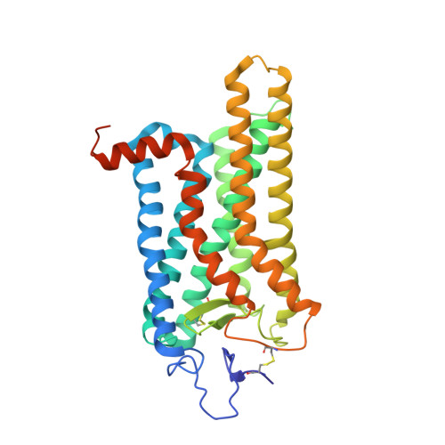
6FK8

Crystal structure of N2C/D282C stabilized opsin bound to RS08


Experimental Data Snapshot

  • Method: X-RAY DIFFRACTION
  • Resolution: 2.87 Å
  • R-Value Free: 0.247 
  • R-Value Work: 0.229 
  • R-Value Observed: 0.229 

Starting Model: experimental
View more details

wwPDB Validation   3D Report Full Report


Ligand Structure Quality Assessment 


This is version 2.2 of the entry. See complete history


Literature

Ligand channel in pharmacologically stabilized rhodopsin.

Mattle, D.Kuhn, B.Aebi, J.Bedoucha, M.Kekilli, D.Grozinger, N.Alker, A.Rudolph, M.G.Schmid, G.Schertler, G.F.X.Hennig, M.Standfuss, J.Dawson, R.J.P.

(2018) Proc Natl Acad Sci U S A 115: 3640-3645

  • DOI: https://doi.org/10.1073/pnas.1718084115
  • Primary Citation of Related Structures:  
    6FK6, 6FK7, 6FK8, 6FK9, 6FKA, 6FKB, 6FKC, 6FKD

  • PubMed Abstract: 

    In the degenerative eye disease retinitis pigmentosa (RP), protein misfolding leads to fatal consequences for cell metabolism and rod and cone cell survival. To stop disease progression, a therapeutic approach focuses on stabilizing inherited protein mutants of the G protein-coupled receptor (GPCR) rhodopsin using pharmacological chaperones (PC) that improve receptor folding and trafficking. In this study, we discovered stabilizing nonretinal small molecules by virtual and thermofluor screening and determined the crystal structure of pharmacologically stabilized opsin at 2.4 Å resolution using one of the stabilizing hits (S-RS1). Chemical modification of S-RS1 and further structural analysis revealed the core binding motif of this class of rhodopsin stabilizers bound at the orthosteric binding site. Furthermore, previously unobserved conformational changes are visible at the intradiscal side of the seven-transmembrane helix bundle. A hallmark of this conformation is an open channel connecting the ligand binding site with the membrane and the intradiscal lumen of rod outer segments. Sufficient in size, the passage permits the exchange of hydrophobic ligands such as retinal. The results broaden our understanding of rhodopsin's conformational flexibility and enable therapeutic drug intervention against rhodopsin-related retinitis pigmentosa.


  • Organizational Affiliation

    Roche Pharma Research and Early Development, Therapeutic Modalities, Roche Innovation Center Basel, F. Hoffmann-La Roche Ltd, 4070 Basel, Switzerland.


Macromolecules
Find similar proteins by:  (by identity cutoff)  |  3D Structure
Entity ID: 1
MoleculeChains Sequence LengthOrganismDetailsImage
Rhodopsin349Bos taurusMutation(s): 1 
Gene Names: RHO
Membrane Entity: Yes 
UniProt
Find proteins for P02699 (Bos taurus)
Explore P02699 
Go to UniProtKB:  P02699
Entity Groups  
Sequence Clusters30% Identity50% Identity70% Identity90% Identity95% Identity100% Identity
UniProt GroupP02699
Glycosylation
Glycosylation Sites: 1
Sequence Annotations
Expand
  • Reference Sequence
Oligosaccharides

Help

Entity ID: 2
MoleculeChains Length2D Diagram Glycosylation3D Interactions
alpha-D-mannopyranose-(1-3)-[alpha-D-mannopyranose-(1-6)]beta-D-mannopyranose-(1-4)-2-acetamido-2-deoxy-beta-D-glucopyranose-(1-4)-2-acetamido-2-deoxy-beta-D-glucopyranose
B
5N-Glycosylation
Glycosylation Resources
GlyTouCan:  G22768VO
GlyCosmos:  G22768VO
GlyGen:  G22768VO
Experimental Data & Validation

Experimental Data

  • Method: X-RAY DIFFRACTION
  • Resolution: 2.87 Å
  • R-Value Free: 0.247 
  • R-Value Work: 0.229 
  • R-Value Observed: 0.229 
  • Space Group: H 3 2
Unit Cell:
Length ( Å )Angle ( ˚ )
a = 242.183α = 90
b = 242.183β = 90
c = 110.967γ = 120
Software Package:
Software NamePurpose
PHENIXrefinement
XDSdata reduction
Aimlessdata scaling

Structure Validation

View Full Validation Report


Ligand Structure Quality Assessment 


Entry History & Funding Information

Deposition Data


Funding OrganizationLocationGrant Number
RocheSwitzerlandRPF

Revision History  (Full details and data files)

  • Version 1.0: 2018-04-04
    Type: Initial release
  • Version 1.1: 2018-04-11
    Changes: Data collection, Database references
  • Version 2.0: 2020-07-29
    Type: Remediation
    Reason: Carbohydrate remediation
    Changes: Atomic model, Data collection, Derived calculations, Refinement description, Structure summary
  • Version 2.1: 2024-01-17
    Changes: Data collection, Database references, Derived calculations, Refinement description, Structure summary
  • Version 2.2: 2024-11-13
    Changes: Structure summary