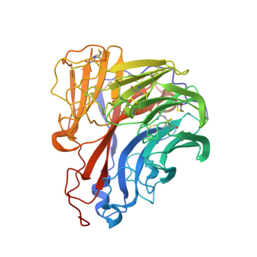
6G02

Complex of neuraminidase from H1N1 influenza virus with tamiphosphor omega-azidohexyl ester


Experimental Data Snapshot

  • Method: X-RAY DIFFRACTION
  • Resolution: 1.84 Å
  • R-Value Free: 0.197 
  • R-Value Work: 0.153 
  • R-Value Observed: 0.154 

wwPDB Validation   3D Report Full Report


Ligand Structure Quality Assessment 


This is version 1.1 of the entry. See complete history


Literature

DNA-linked inhibitor antibody assay (DIANA) as a new method for screening influenza neuraminidase inhibitors.

Kozisek, M.Navratil, V.Rojikova, K.Pokorna, J.Berenguer Albinana, C.Pachl, P.Zemanova, J.Machara, A.Sacha, P.Hudlicky, J.Cisarova, I.Rezacova, P.Konvalinka, J.

(2018) Biochem J 475: 3847-3860

  • DOI: https://doi.org/10.1042/BCJ20180764
  • Primary Citation of Related Structures:  
    6G01, 6G02

  • PubMed Abstract: 

    Influenza neuraminidase is responsible for the escape of new viral particles from the infected cell surface. Several neuraminidase inhibitors are used clinically to treat patients or stockpiled for emergencies. However, the increasing development of viral resistance against approved inhibitors has underscored the need for the development of new antivirals effective against resistant influenza strains. A facile, sensitive, and inexpensive screening method would help achieve this goal. Recently, we described a multiwell plate-based DNA-linked inhibitor antibody assay (DIANA). This highly sensitive method can quantify femtomolar concentrations of enzymes. DIANA also has been applied to high-throughput enzyme inhibitor screening, allowing the evaluation of inhibition constants from a single inhibitor concentration. Here, we report the design, synthesis, and structural characterization of a tamiphosphor derivative linked to a reporter DNA oligonucleotide for the development of a DIANA-type assay to screen potential influenza neuraminidase inhibitors. The neuraminidase is first captured by an immobilized antibody, and the test compound competes for binding to the enzyme with the oligo-linked detection probe, which is then quantified by qPCR. We validated this novel assay by comparing it with the standard fluorometric assay and demonstrated its usefulness for sensitive neuraminidase detection as well as high-throughput screening of potential new neuraminidase inhibitors.


  • Organizational Affiliation

    Institute of Organic Chemistry and Biochemistry of the Czech Academy of Sciences, Gilead Sciences and IOCB Research Center, Flemingovo n. 2, 16610 Prague 6, Czech Republic milan.kozisek@uochb.cas.cz konval@uochb.cas.cz.


Macromolecules
Find similar proteins by:  (by identity cutoff)  |  3D Structure
Entity ID: 1
MoleculeChains Sequence LengthOrganismDetailsImage
Neuraminidase
A, B
387Influenza A virus (A/California/07/2009(H1N1))Mutation(s): 0 
Gene Names: NA
EC: 3.2.1.18
UniProt
Find proteins for C3W6G3 (Influenza A virus)
Explore C3W6G3 
Go to UniProtKB:  C3W6G3
Entity Groups  
Sequence Clusters30% Identity50% Identity70% Identity90% Identity95% Identity100% Identity
UniProt GroupC3W6G3
Glycosylation
Glycosylation Sites: 3
Sequence Annotations
Expand
  • Reference Sequence
Small Molecules
Ligands 4 Unique
IDChains Name / Formula / InChI Key2D Diagram3D Interactions
EF8
Query on EF8

Download Ideal Coordinates CCD File 
D [auth A],
P [auth B]
6-[[(3~{R},4~{R},5~{S})-4-acetamido-5-azanyl-3-pentan-3-yloxy-cyclohexen-1-yl]-oxidanyl-phosphoryl]oxyhexylimino-azanylidene-azanium
C19 H37 N5 O5 P
SQYBSQHYIJSZEH-IPMKNSEASA-O
NAG
Query on NAG

Download Ideal Coordinates CCD File 
E [auth A]
F [auth A]
G [auth A]
Q [auth B]
R [auth B]
E [auth A],
F [auth A],
G [auth A],
Q [auth B],
R [auth B],
S [auth B]
2-acetamido-2-deoxy-beta-D-glucopyranose
C8 H15 N O6
OVRNDRQMDRJTHS-FMDGEEDCSA-N
EDO
Query on EDO

Download Ideal Coordinates CCD File 
H [auth A]
I [auth A]
J [auth A]
K [auth A]
L [auth A]
H [auth A],
I [auth A],
J [auth A],
K [auth A],
L [auth A],
M [auth A],
T [auth B],
U [auth B],
V [auth B],
W [auth B],
X [auth B]
1,2-ETHANEDIOL
C2 H6 O2
LYCAIKOWRPUZTN-UHFFFAOYSA-N
CA
Query on CA

Download Ideal Coordinates CCD File 
C [auth A],
N [auth B],
O [auth B]
CALCIUM ION
Ca
BHPQYMZQTOCNFJ-UHFFFAOYSA-N
Experimental Data & Validation

Experimental Data

  • Method: X-RAY DIFFRACTION
  • Resolution: 1.84 Å
  • R-Value Free: 0.197 
  • R-Value Work: 0.153 
  • R-Value Observed: 0.154 
  • Space Group: P 4
Unit Cell:
Length ( Å )Angle ( ˚ )
a = 114.86α = 90
b = 114.86β = 90
c = 63.899γ = 90
Software Package:
Software NamePurpose
REFMACrefinement
XDSdata reduction
MOLREPphasing
PDB_EXTRACTdata extraction
XDSdata scaling

Structure Validation

View Full Validation Report


Ligand Structure Quality Assessment 


Entry History & Funding Information

Deposition Data


Funding OrganizationLocationGrant Number
Ministry of Education, Youth and Sports of the Czech RepublicCzech RepublicInterBioMed LO1302

Revision History  (Full details and data files)

  • Version 1.0: 2019-01-23
    Type: Initial release
  • Version 1.1: 2020-07-29
    Type: Remediation
    Reason: Carbohydrate remediation
    Changes: Data collection, Derived calculations, Structure summary