6JS8

Structure of the CYP102A1 Haem Domain with N-Dehydroabietoyl-L-Tryptophan


Experimental Data Snapshot

  • Method: X-RAY DIFFRACTION
  • Resolution: 1.36 Å
  • R-Value Free: 
    0.159 (Depositor), 0.160 (DCC) 
  • R-Value Work: 
    0.132 (Depositor), 0.130 (DCC) 
  • R-Value Observed: 
    0.133 (Depositor) 

Starting Model: experimental
View more details

wwPDB Validation   3D Report Full Report


Ligand Structure Quality Assessment 

Created with Raphaël 2.3.0Worse 01 BetterLigand structure goodness of fit to experimental dataBest fitted C5RClick on this verticalbar to view details

This is version 1.3 of the entry. See complete history


Literature

Crystals in Minutes: Instant On-Site Microcrystallisation of Various Flavours of the CYP102A1 (P450BM3) Haem Domain.

Stanfield, J.K.Omura, K.Matsumoto, A.Kasai, C.Sugimoto, H.Shiro, Y.Watanabe, Y.Shoji, O.

(2020) Angew Chem Int Ed Engl 59: 7611-7618

  • DOI: https://doi.org/10.1002/anie.201913407
  • Primary Citation of Related Structures:  
    6JLV, 6JMH, 6JMW, 6JO1, 6JS8, 6JVC, 6JZS, 6K24, 6K58, 6K9S

  • PubMed Abstract: 

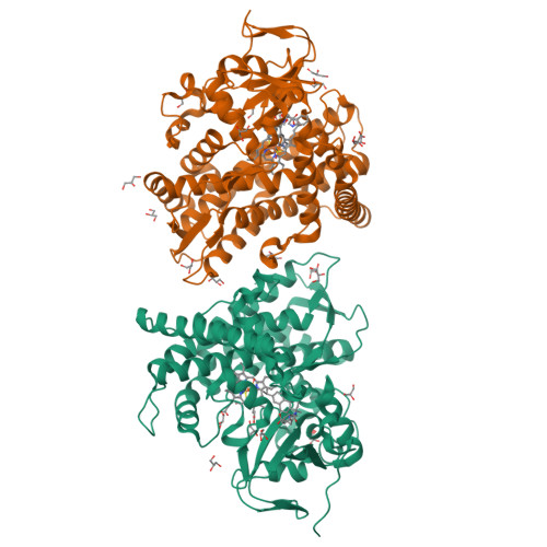
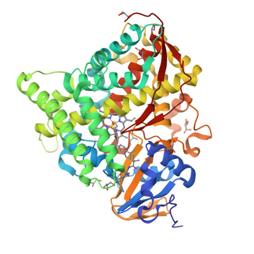
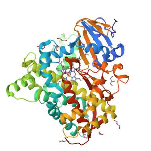
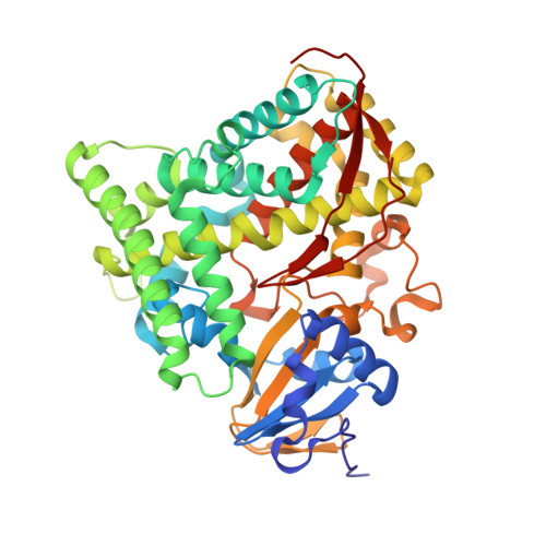
    Despite CYP102A1 (P450BM3) representing one of the most extensively researched metalloenzymes, crystallisation of its haem domain upon modification can be a challenge. Crystal structures are indispensable for the efficient structure-based design of P450BM3 as a biocatalyst. The abietane diterpenoid derivative N-abietoyl-l-tryptophan (AbiATrp) is an outstanding crystallisation accelerator for the wild-type P450BM3 haem domain, with visible crystals forming within 2 hours and diffracting to a near-atomic resolution of 1.22 Å. Using these crystals as seeds in a cross-microseeding approach, an assortment of P450BM3 haem domain crystal structures, containing previously uncrystallisable decoy molecules and diverse artificial metalloporphyrins binding various ligand molecules, as well as heavily tagged haem-domain variants, could be determined. Some of the structures reported herein could be used as models of different stages of the P450BM3 catalytic cycle.


  • Organizational Affiliation

    Department of Chemistry, Graduate School of Science, Nagoya University, Furo-cho, Chikusa-ku, Nagoya, 464-8602, Japan.


Macromolecules
Find similar proteins by:  (by identity cutoff)  |  3D Structure
Entity ID: 1
MoleculeChains Sequence LengthOrganismDetailsImage
Bifunctional cytochrome P450/NADPH--P450 reductase
A, B
456Priestia megateriumMutation(s): 0 
Gene Names: cyp102A1
EC: 1.14.14.1 (PDB Primary Data), 1.6.2.4 (PDB Primary Data)
UniProt
Find proteins for P14779 (Priestia megaterium (strain ATCC 14581 / DSM 32 / CCUG 1817 / JCM 2506 / NBRC 15308 / NCIMB 9376 / NCTC 10342 / NRRL B-14308 / VKM B-512 / Ford 19))
Explore P14779 
Go to UniProtKB:  P14779
Entity Groups  
Sequence Clusters30% Identity50% Identity70% Identity90% Identity95% Identity100% Identity
UniProt GroupP14779
Sequence Annotations
Expand
  • Reference Sequence
Small Molecules
Ligands 4 Unique
IDChains Name / Formula / InChI Key2D Diagram3D Interactions
HEM
Query on HEM

Download Ideal Coordinates CCD File 
C [auth A],
O [auth B]
PROTOPORPHYRIN IX CONTAINING FE
C34 H32 Fe N4 O4
KABFMIBPWCXCRK-RGGAHWMASA-L
C5R (Subject of Investigation/LOI)
Query on C5R

Download Ideal Coordinates CCD File 
E [auth A],
Q [auth B]
(2S)-2-[[(1R,4aS,10aR)-1,4a-dimethyl-7-propan-2-yl-2,3,4,9,10,10a-hexahydrophenanthren-1-yl]carbonylamino]-3-(1H-indol-3-yl)propanoic acid
C31 H38 N2 O3
WYAQKAIGBDDJPR-QKOWHPQASA-N
GOL
Query on GOL

Download Ideal Coordinates CCD File 
AA [auth B]
BA [auth B]
F [auth A]
G [auth A]
H [auth A]
GLYCEROL
C3 H8 O3
PEDCQBHIVMGVHV-UHFFFAOYSA-N
DMS
Query on DMS

Download Ideal Coordinates CCD File 
D [auth A],
P [auth B]
DIMETHYL SULFOXIDE
C2 H6 O S
IAZDPXIOMUYVGZ-UHFFFAOYSA-N
Experimental Data & Validation

Experimental Data

  • Method: X-RAY DIFFRACTION
  • Resolution: 1.36 Å
  • R-Value Free:  0.159 (Depositor), 0.160 (DCC) 
  • R-Value Work:  0.132 (Depositor), 0.130 (DCC) 
  • R-Value Observed: 0.133 (Depositor) 
Space Group: P 1 21 1
Unit Cell:
Length ( Å )Angle ( ˚ )
a = 58.886α = 90
b = 148.789β = 98.42
c = 63.196γ = 90
Software Package:
Software NamePurpose
XDSdata reduction
Aimlessdata scaling
MOLREPphasing
REFMACrefinement
PDB_EXTRACTdata extraction

Structure Validation

View Full Validation Report


Ligand Structure Quality Assessment 

Created with Raphaël 2.3.0Worse 01 BetterLigand structure goodness of fit to experimental dataBest fitted C5RClick on this verticalbar to view details

Entry History & Funding Information

Deposition Data


Funding OrganizationLocationGrant Number
Japan Science and TechnologyJapanJPMJCR15P3

Revision History  (Full details and data files)

  • Version 1.0: 2020-03-18
    Type: Initial release
  • Version 1.1: 2020-03-25
    Changes: Database references
  • Version 1.2: 2020-05-06
    Changes: Database references
  • Version 1.3: 2023-11-22
    Changes: Data collection, Database references, Derived calculations, Refinement description