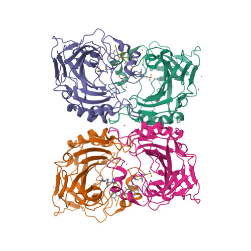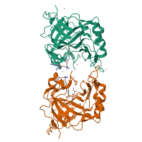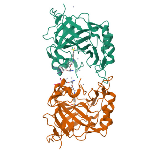
PROTEIN-AROMATIC FOLDAMER COMPLEX CRYSTAL STRUCTURE
Zeberko, C.To be published.
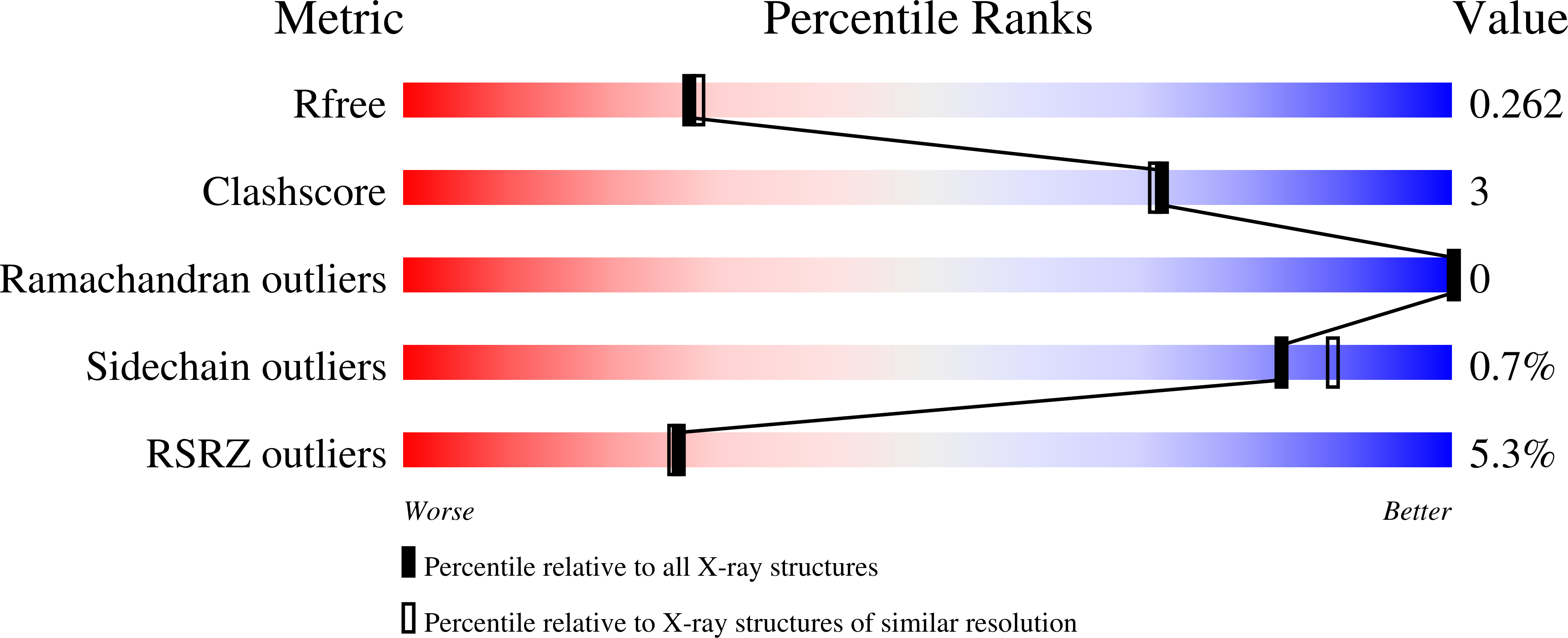
Experimental Data Snapshot
Starting Model: experimental
View more details
Entity ID: 1 | |||||
|---|---|---|---|---|---|
| Molecule | Chains | Sequence Length | Organism | Details | Image |
| Carbonic anhydrase 2 | 259 | Homo sapiens | Mutation(s): 0 Gene Names: CA2 EC: 4.2.1.1 (PDB Primary Data), 4.2.1.69 (UniProt) | 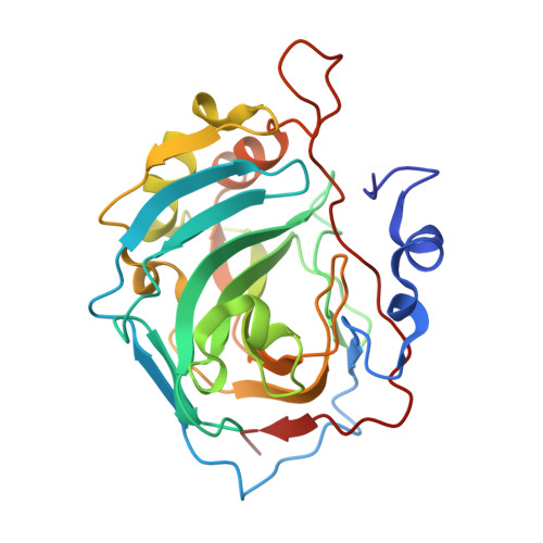 | |
UniProt & NIH Common Fund Data Resources | |||||
Find proteins for P00918 (Homo sapiens) Explore P00918 Go to UniProtKB: P00918 | |||||
PHAROS: P00918 GTEx: ENSG00000104267 | |||||
Entity Groups | |||||
| Sequence Clusters | 30% Identity50% Identity70% Identity90% Identity95% Identity100% Identity | ||||
| UniProt Group | P00918 | ||||
Sequence AnnotationsExpand | |||||
| |||||
Find similar proteins by: Sequence | 3D Structure
Entity ID: 2 | |||||
|---|---|---|---|---|---|
| Molecule | Chains | Sequence Length | Organism | Details | Image |
| Aromatic foldamer | E [auth H], F [auth E], G [auth F], H [auth G] | 5 | synthetic construct | Mutation(s): 0 | 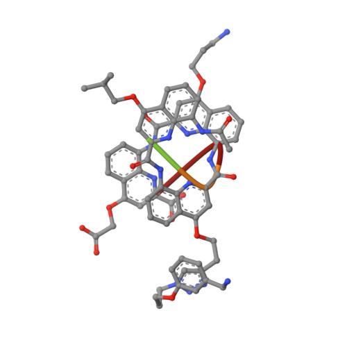 |
Sequence AnnotationsExpand | |||||
| |||||
| Ligands 3 Unique | |||||
|---|---|---|---|---|---|
| ID | Chains | Name / Formula / InChI Key | 2D Diagram | 3D Interactions | |
| 4SO Query on 4SO | MA [auth H], NA [auth E], OA [auth F], PA [auth G] | 4-sulfamoylbenzoic acid C7 H7 N O4 S UCAGLBKTLXCODC-UHFFFAOYSA-N |  | ||
| GOL Query on GOL | DA [auth C] | GLYCEROL C3 H8 O3 PEDCQBHIVMGVHV-UHFFFAOYSA-N |  | ||
| ZN Query on ZN | AA [auth C] BA [auth C] CA [auth C] EA [auth C] FA [auth C] | ZINC ION Zn PTFCDOFLOPIGGS-UHFFFAOYSA-N |  | ||
| Length ( Å ) | Angle ( ˚ ) |
|---|---|
| a = 59.106 | α = 66.1 |
| b = 76.478 | β = 86.83 |
| c = 81.417 | γ = 73.6 |
| Software Name | Purpose |
|---|---|
| REFMAC | refinement |
| CrysalisPro | data reduction |
| Aimless | data scaling |
| MOLREP | phasing |
RCSB PDB is hosted by

RCSB PDB is a member of the