6QDJ | pdb_00006qdj

Molecular features of the UNC-45 chaperone critical for binding and folding muscle myosin


Experimental Data Snapshot

  • Method: X-RAY DIFFRACTION
  • Resolution: 1.88 Å
  • R-Value Free: 
    0.213 (Depositor), 0.220 (DCC) 
  • R-Value Work: 
    0.184 (Depositor), 0.190 (DCC) 
  • R-Value Observed: 
    0.186 (Depositor) 

Starting Model: experimental
View more details

wwPDB Validation   3D Report Full Report


Ligand Structure Quality Assessment 

Created with Raphaël 2.3.0Worse 01 BetterLigand structure goodness of fit to experimental dataBest fitted ADPClick on this verticalbar to view details

This is version 1.1 of the entry. See complete history


Literature

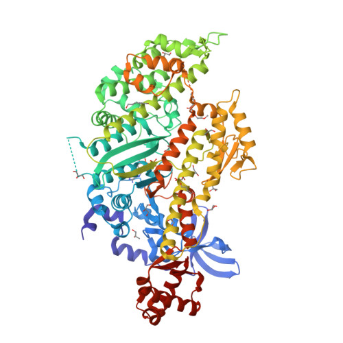
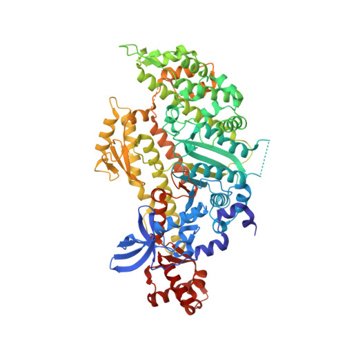
Molecular features of the UNC-45 chaperone critical for binding and folding muscle myosin.

Hellerschmied, D.Lehner, A.Franicevic, N.Arnese, R.Johnson, C.Vogel, A.Meinhart, A.Kurzbauer, R.Deszcz, L.Gazda, L.Geeves, M.Clausen, T.

(2019) Nat Commun 10: 4781-4781

  • DOI: https://doi.org/10.1038/s41467-019-12667-8
  • Primary Citation of Related Structures:  
    6QDJ, 6QDK, 6QDL, 6QDM

  • PubMed Abstract: 

    Myosin is a motor protein that is essential for a variety of processes ranging from intracellular transport to muscle contraction. Folding and assembly of myosin relies on a specific chaperone, UNC-45. To address its substrate-targeting mechanism, we reconstitute the interplay between Caenorhabditis elegans UNC-45 and muscle myosin MHC-B in insect cells. In addition to providing a cellular chaperone assay, the established system enabled us to produce large amounts of functional muscle myosin, as evidenced by a biochemical and structural characterization, and to directly monitor substrate binding to UNC-45. Data from in vitro and cellular chaperone assays, together with crystal structures of binding-deficient UNC-45 mutants, highlight the importance of utilizing a flexible myosin-binding domain. This so-called UCS domain can adopt discrete conformations to efficiently bind and fold substrate. Moreover, our data uncover the molecular basis of temperature-sensitive UNC-45 mutations underlying one of the most prominent motility defects in C. elegans.


  • Organizational Affiliation

    Research Institute of Molecular Pathology, Vienna BioCenter, Vienna, Austria. doris.hellerschmied@uni-due.de.


Macromolecules
Find similar proteins by:  (by identity cutoff)  |  3D Structure
Entity ID: 1
MoleculeChains Sequence LengthOrganismDetailsImage
Myosin-4790Caenorhabditis elegansMutation(s): 0 
Gene Names: unc-54myo-4F11C3.3
UniProt
Find proteins for P02566 (Caenorhabditis elegans)
Explore P02566 
Go to UniProtKB:  P02566
Entity Groups  
Sequence Clusters30% Identity50% Identity70% Identity90% Identity95% Identity100% Identity
UniProt GroupP02566
Sequence Annotations
Expand
  • Reference Sequence
Small Molecules
Ligands 6 Unique
IDChains Name / Formula / InChI Key2D Diagram3D Interactions
ADP
Query on ADP

Download Ideal Coordinates CCD File 
B [auth A]ADENOSINE-5'-DIPHOSPHATE
C10 H15 N5 O10 P2
XTWYTFMLZFPYCI-KQYNXXCUSA-N
P15
Query on P15

Download Ideal Coordinates CCD File 
Q [auth A]2,5,8,11,14,17-HEXAOXANONADECAN-19-OL
C13 H28 O7
FHHGCKHKTAJLOM-UHFFFAOYSA-N
HEZ
Query on HEZ

Download Ideal Coordinates CCD File 
P [auth A]HEXANE-1,6-DIOL
C6 H14 O2
XXMIOPMDWAUFGU-UHFFFAOYSA-N
GOL
Query on GOL

Download Ideal Coordinates CCD File 
C [auth A]
D [auth A]
E [auth A]
F [auth A]
G [auth A]
GLYCEROL
C3 H8 O3
PEDCQBHIVMGVHV-UHFFFAOYSA-N
BU1
Query on BU1

Download Ideal Coordinates CCD File 
M [auth A]1,4-BUTANEDIOL
C4 H10 O2
WERYXYBDKMZEQL-UHFFFAOYSA-N
PGO
Query on PGO

Download Ideal Coordinates CCD File 
N [auth A],
O [auth A]
S-1,2-PROPANEDIOL
C3 H8 O2
DNIAPMSPPWPWGF-VKHMYHEASA-N
Experimental Data & Validation

Experimental Data

  • Method: X-RAY DIFFRACTION
  • Resolution: 1.88 Å
  • R-Value Free:  0.213 (Depositor), 0.220 (DCC) 
  • R-Value Work:  0.184 (Depositor), 0.190 (DCC) 
  • R-Value Observed: 0.186 (Depositor) 
Space Group: P 1 21 1
Unit Cell:
Length ( Å )Angle ( ˚ )
a = 54.11α = 90
b = 111.96β = 97.44
c = 84.74γ = 90
Software Package:
Software NamePurpose
PHENIXrefinement
Cootmodel building
PHENIXmodel building
XDSdata reduction
XSCALEdata scaling
PHASERphasing

Structure Validation

View Full Validation Report


Ligand Structure Quality Assessment 

Created with Raphaël 2.3.0Worse 01 BetterLigand structure goodness of fit to experimental dataBest fitted ADPClick on this verticalbar to view details

Entry History & Funding Information

Deposition Data


Funding OrganizationLocationGrant Number
Austrian Science FundAustriaP 22570

Revision History  (Full details and data files)

  • Version 1.0: 2019-10-30
    Type: Initial release
  • Version 1.1: 2024-01-24
    Changes: Data collection, Database references, Refinement description