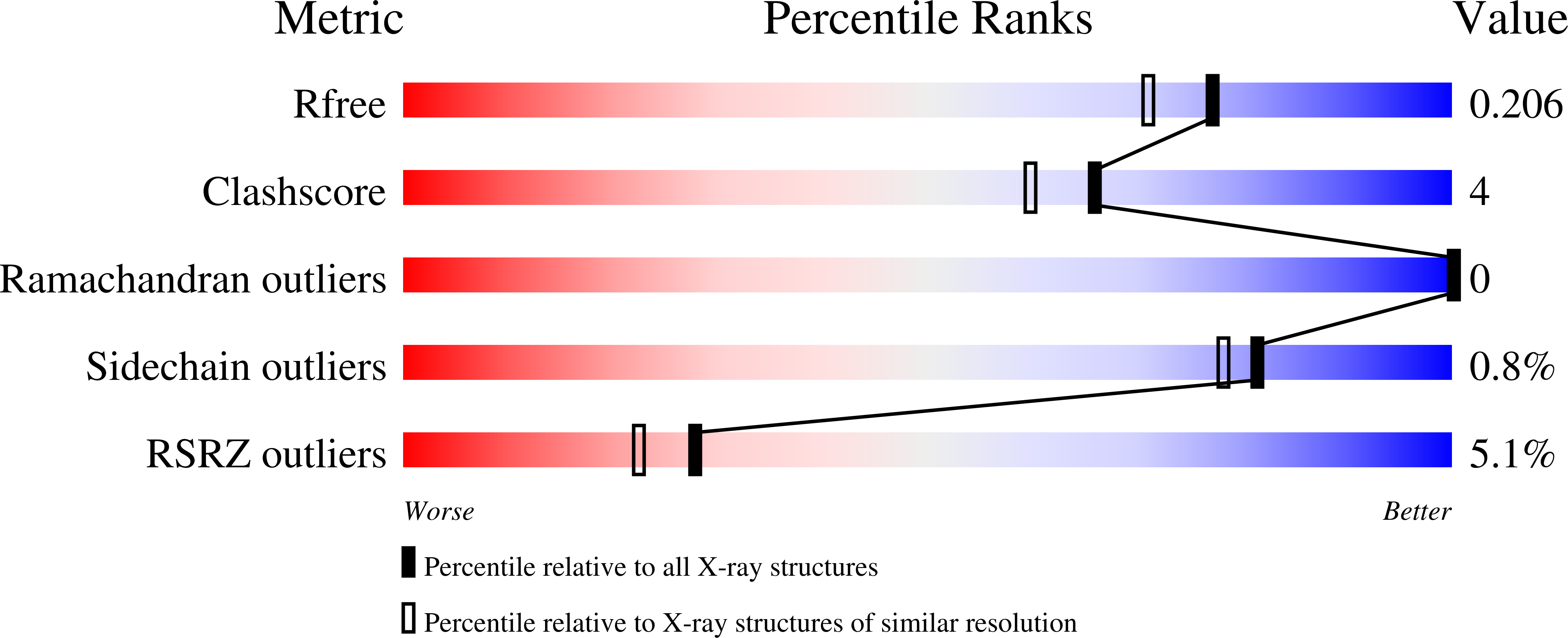
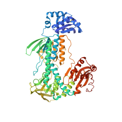
6ZP3

Thioredoxin glutathione reductase from Schistosoma mansoni in complex with 2-Methylindole-3-acetic acid


Experimental Data Snapshot

  • Method: X-RAY DIFFRACTION
  • Resolution: 1.80 Å
  • R-Value Free: 0.205 
  • R-Value Work: 0.172 
  • R-Value Observed: 0.174 

Starting Model: experimental
View more details

wwPDB Validation   3D Report Full Report


Ligand Structure Quality Assessment 


This is version 1.3 of the entry. See complete history


Literature

Probing the Surface of a Parasite Drug Target Thioredoxin Glutathione Reductase Using Small Molecule Fragments.

Fata, F.Silvestri, I.Ardini, M.Ippoliti, R.Di Leandro, L.Demitri, N.Polentarutti, M.Di Matteo, A.Lyu, H.Thatcher, G.R.J.Petukhov, P.A.Williams, D.L.Angelucci, F.

(2021) ACS Infect Dis 7: 1932-1944

  • DOI: https://doi.org/10.1021/acsinfecdis.0c00909
  • Primary Citation of Related Structures:  
    6ZLB, 6ZLP, 6ZP3, 6ZST, 7B02, 7NPX

  • PubMed Abstract: 

    Fragment screening is a powerful drug discovery approach particularly useful for enzymes difficult to inhibit selectively, such as the thiol/selenol-dependent thioredoxin reductases (TrxRs), which are essential and druggable in several infectious diseases. Several known inhibitors are reactive electrophiles targeting the selenocysteine-containing C-terminus and thus often suffering from off-target reactivity in vivo . The lack of structural information on the interaction modalities of the C-terminus-targeting inhibitors, due to the high mobility of this domain and the lack of alternative druggable sites, prevents the development of selective inhibitors for TrxRs. In this work, fragments selected from actives identified in a large screen carried out against Thioredoxin Glutathione Reductase from Schistosoma mansoni (SmTGR) were probed by X-ray crystallography. SmTGR is one of the most promising drug targets for schistosomiasis, a devastating, neglected disease. Utilizing a multicrystal method to analyze electron density maps, structural analysis, and functional studies, three binding sites were characterized in SmTGR: two sites are close to or partially superposable with the NADPH binding site, while the third one is found between two symmetry related SmTGR subunits of the crystal lattice. Surprisingly, one compound bound to this latter site stabilizes, through allosteric effects mediated by the so-called guiding bar residues, the crucial redox active C-terminus of SmTGR, making it finally visible at high resolution. These results further promote fragments as small molecule probes for investigating functional aspects of the target protein, exemplified by the allosteric effect on the C-terminus, and providing fundamental chemical information exploitable in drug discovery.


  • Organizational Affiliation

    Department of Life, Health and Environmental Sciences, University of L'Aquila, 67100 L'Aquila, Italy.


Macromolecules
Find similar proteins by:  (by identity cutoff)  |  3D Structure
Entity ID: 1
MoleculeChains Sequence LengthOrganismDetailsImage
Thioredoxin glutathione reductase598Schistosoma mansoniMutation(s): 0 
Gene Names: Smp_048430
EC: 1.8.1.9
UniProt
Find proteins for A0A3Q0KFL1 (Schistosoma mansoni)
Explore A0A3Q0KFL1 
Go to UniProtKB:  A0A3Q0KFL1
Entity Groups  
Sequence Clusters30% Identity50% Identity70% Identity90% Identity95% Identity100% Identity
UniProt GroupA0A3Q0KFL1
Sequence Annotations
Expand
  • Reference Sequence
Small Molecules
Ligands 9 Unique
IDChains Name / Formula / InChI Key2D Diagram3D Interactions
FAD
Query on FAD

Download Ideal Coordinates CCD File 
B [auth A]FLAVIN-ADENINE DINUCLEOTIDE
C27 H33 N9 O15 P2
VWWQXMAJTJZDQX-UYBVJOGSSA-N
PG4
Query on PG4

Download Ideal Coordinates CCD File 
D [auth A]TETRAETHYLENE GLYCOL
C8 H18 O5
UWHCKJMYHZGTIT-UHFFFAOYSA-N
QN5 (Subject of Investigation/LOI)
Query on QN5

Download Ideal Coordinates CCD File 
F [auth A]2-(2-methyl-1~{H}-indol-3-yl)ethanoic acid
C11 H11 N O2
QJNNHJVSQUUHHE-UHFFFAOYSA-N
DTT
Query on DTT

Download Ideal Coordinates CCD File 
I [auth A]2,3-DIHYDROXY-1,4-DITHIOBUTANE
C4 H10 O2 S2
VHJLVAABSRFDPM-IMJSIDKUSA-N
PGE
Query on PGE

Download Ideal Coordinates CCD File 
J [auth A]TRIETHYLENE GLYCOL
C6 H14 O4
ZIBGPFATKBEMQZ-UHFFFAOYSA-N
PEG
Query on PEG

Download Ideal Coordinates CCD File 
H [auth A]DI(HYDROXYETHYL)ETHER
C4 H10 O3
MTHSVFCYNBDYFN-UHFFFAOYSA-N
DMS
Query on DMS

Download Ideal Coordinates CCD File 
E [auth A]DIMETHYL SULFOXIDE
C2 H6 O S
IAZDPXIOMUYVGZ-UHFFFAOYSA-N
CA
Query on CA

Download Ideal Coordinates CCD File 
G [auth A]CALCIUM ION
Ca
BHPQYMZQTOCNFJ-UHFFFAOYSA-N
K
Query on K

Download Ideal Coordinates CCD File 
C [auth A]POTASSIUM ION
K
NPYPAHLBTDXSSS-UHFFFAOYSA-N
Experimental Data & Validation

Experimental Data

  • Method: X-RAY DIFFRACTION
  • Resolution: 1.80 Å
  • R-Value Free: 0.205 
  • R-Value Work: 0.172 
  • R-Value Observed: 0.174 
  • Space Group: C 1 2 1
Unit Cell:
Length ( Å )Angle ( ˚ )
a = 141.849α = 90
b = 102.186β = 112.94
c = 58.572γ = 90
Software Package:
Software NamePurpose
PHENIXrefinement
PDB_EXTRACTdata extraction
XDSdata reduction
Aimlessdata scaling
PHASERphasing

Structure Validation

View Full Validation Report


Ligand Structure Quality Assessment 


Entry History & Funding Information

Deposition Data


Funding OrganizationLocationGrant Number
National Institutes of Health/National Institute Of Allergy and Infectious Diseases (NIH/NIAID)United StatesR33AI127635

Revision History  (Full details and data files)

  • Version 1.0: 2021-05-19
    Type: Initial release
  • Version 1.1: 2021-07-21
    Changes: Database references
  • Version 1.2: 2024-01-31
    Changes: Data collection, Database references, Refinement description
  • Version 1.3: 2024-11-20
    Changes: Structure summary