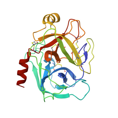
Room-temperature crystallography using a microfluidic protein crystal array device and its application to protein-ligand complex structure analysis.
Maeki, M., Ito, S., Takeda, R., Ueno, G., Ishida, A., Tani, H., Yamamoto, M., Tokeshi, M.(2020) Chem Sci 11: 9072-9087
- PubMed: 34094189
- DOI: https://doi.org/10.1039/d0sc02117b
- Primary Citation of Related Structures:
7BRV, 7BRW, 7BRX, 7BRY, 7BRZ, 7BS0, 7BS1, 7BS2, 7BS3, 7BS4, 7BS5, 7BS6, 7BS7, 7BS8, 7BS9, 7BSA - PubMed Abstract:
Room-temperature (RT) protein crystallography provides significant information to elucidate protein function under physiological conditions. In particular, contrary to typical binding assays, X-ray crystal structure analysis of a protein-ligand complex can determine the three-dimensional (3D) configuration of its binding site. This allows the development of effective drugs by structure-based and fragment-based (FBDD) drug design. However, RT crystallography and RT crystallography-based protein-ligand complex analyses require the preparation and measurement of numerous crystals to avoid the X-ray radiation damage. Thus, for the application of RT crystallography to protein-ligand complex analysis, the simultaneous preparation of protein-ligand complex crystals and sequential X-ray diffraction measurement remain challenging. Here, we report an RT crystallography technique using a microfluidic protein crystal array device for protein-ligand complex structure analysis. We demonstrate the microfluidic sorting of protein crystals into microwells without any complicated procedures and apparatus, whereby the sorted protein crystals are fixed into microwells and sequentially measured to collect X-ray diffraction data. This is followed by automatic data processing to calculate the 3D protein structure. The microfluidic device allows the high-throughput preparation of the protein-ligand complex solely by the replacement of the microchannel content with the required ligand solution. We determined eight trypsin-ligand complex structures for the proof of concept experiment and found differences in the ligand coordination of the corresponding RT and conventional cryogenic structures. This methodology can be applied to easily obtain more natural structures. Moreover, drug development by FBDD could be more effective using the proposed methodology.
Organizational Affiliation:
Division of Applied Chemistry, Faculty of Engineering, Hokkaido University Kita 13 Nishi 8, Kita-ku Sapporo 060-8628 Japan m.maeki@eng.hokudai.ac.jp tokeshi@eng.hokudai.ac.jp +81-11-706-6745 +81-11-706-6745 +81-11-706-6744.


