7P4G

Rabbit Muscle L-lactate dehydrogenase in complex with citrate


Experimental Data Snapshot

  • Method: X-RAY DIFFRACTION
  • Resolution: 2.60 Å
  • R-Value Free: 0.260 
  • R-Value Work: 0.199 
  • R-Value Observed: 0.202 

wwPDB Validation   3D Report Full Report


Ligand Structure Quality Assessment 


This is version 1.1 of the entry. See complete history


Literature

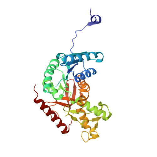
Allosteric transitions of rabbit skeletal muscle lactate dehydrogenase induced by pH-dependent dissociation of the tetrameric enzyme.

Iacovino, L.G.Rossi, M.Di Stefano, G.Rossi, V.Binda, C.Brigotti, M.Tomaselli, F.Pasti, A.P.Dal Piaz, F.Cerini, S.Hochkoeppler, A.

(2022) Biochimie 199: 23-35

  • DOI: https://doi.org/10.1016/j.biochi.2022.03.008
  • Primary Citation of Related Structures:  
    7P4G

  • PubMed Abstract: 

    Among the functions exerted by eukaryotic lactate dehydrogenases, it is of importance the generation of lactate in muscles subjected to fatigue or to limited oxygen availability, with both these conditions triggering a decrease of cellular pH. However, the mutual dependence between lactate dehydrogenase (LDH) catalytic action and lactic acidosis is far from being fully understood. Here we show that the tetrameric LDH from rabbit skeletal muscle undergoes allosteric transitions as a function of pH, i.e. the enzyme obeys Michaelis-Menten kinetics at neutral or slightly alkaline pH values, and features sigmoidal kinetics at pH 6.5 or lower. Remarkably, we also report that a significant dissociation of tetrameric rabbit LDH occurs under acidic conditions, with pyruvate/NAD + or citrate counteracting this effect. Moreover, citrate strongly activates rabbit LDH, inducing the enzyme to feature Michaelis-Menten kinetics. Further, using primary rabbit skeletal muscle cells we tested the generation of lactate as a function of pH, and we detected a parallel decrease of cytosolic pH and secretion of lactate. Overall, our observations indicate that lactic acidosis is antagonized by LDH dissociation, the occurrence of which is regulated by citrate and by allosteric transitions of the enzyme induced by pyruvate.


  • Organizational Affiliation

    Department of Biology and Biotechnology, University of Pavia, Via Ferrata 1, 27100, Pavia, Italy.


Macromolecules
Find similar proteins by:  (by identity cutoff)  |  3D Structure
Entity ID: 1
MoleculeChains Sequence LengthOrganismDetailsImage
L-lactate dehydrogenase A chain
A, B, C, D, E
A, B, C, D, E, F, G, H, I, J, K, L, M, N, O, P
332Oryctolagus cuniculusMutation(s): 1 
Gene Names: LDHA
EC: 1.1.1.27
UniProt
Find proteins for P13491 (Oryctolagus cuniculus)
Explore P13491 
Go to UniProtKB:  P13491
Entity Groups  
Sequence Clusters30% Identity50% Identity70% Identity90% Identity95% Identity100% Identity
UniProt GroupP13491
Sequence Annotations
Expand
  • Reference Sequence
Small Molecules
Ligands 1 Unique
IDChains Name / Formula / InChI Key2D Diagram3D Interactions
CIT (Subject of Investigation/LOI)
Query on CIT

Download Ideal Coordinates CCD File 
AA [auth K]
BA [auth L]
CA [auth M]
DA [auth N]
EA [auth O]
AA [auth K],
BA [auth L],
CA [auth M],
DA [auth N],
EA [auth O],
FA [auth P],
Q [auth A],
R [auth B],
S [auth C],
T [auth D],
U [auth E],
V [auth F],
W [auth G],
X [auth H],
Y [auth I],
Z [auth J]
CITRIC ACID
C6 H8 O7
KRKNYBCHXYNGOX-UHFFFAOYSA-N
Experimental Data & Validation

Experimental Data

  • Method: X-RAY DIFFRACTION
  • Resolution: 2.60 Å
  • R-Value Free: 0.260 
  • R-Value Work: 0.199 
  • R-Value Observed: 0.202 
  • Space Group: P 1
Unit Cell:
Length ( Å )Angle ( ˚ )
a = 83.705α = 114.55
b = 141.571β = 94.69
c = 148.136γ = 102.2
Software Package:
Software NamePurpose
REFMACrefinement
PDB_EXTRACTdata extraction
XDSdata reduction
Aimlessdata scaling
MOLREPphasing

Structure Validation

View Full Validation Report


Ligand Structure Quality Assessment 


Entry History 

Deposition Data

Revision History  (Full details and data files)

  • Version 1.0: 2022-05-18
    Type: Initial release
  • Version 1.1: 2024-01-31
    Changes: Data collection, Refinement description