7RPZ

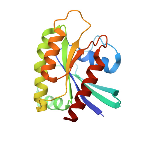
KRAS G12D in complex with MRTX-1133


Experimental Data Snapshot

  • Method: X-RAY DIFFRACTION
  • Resolution: 1.30 Å
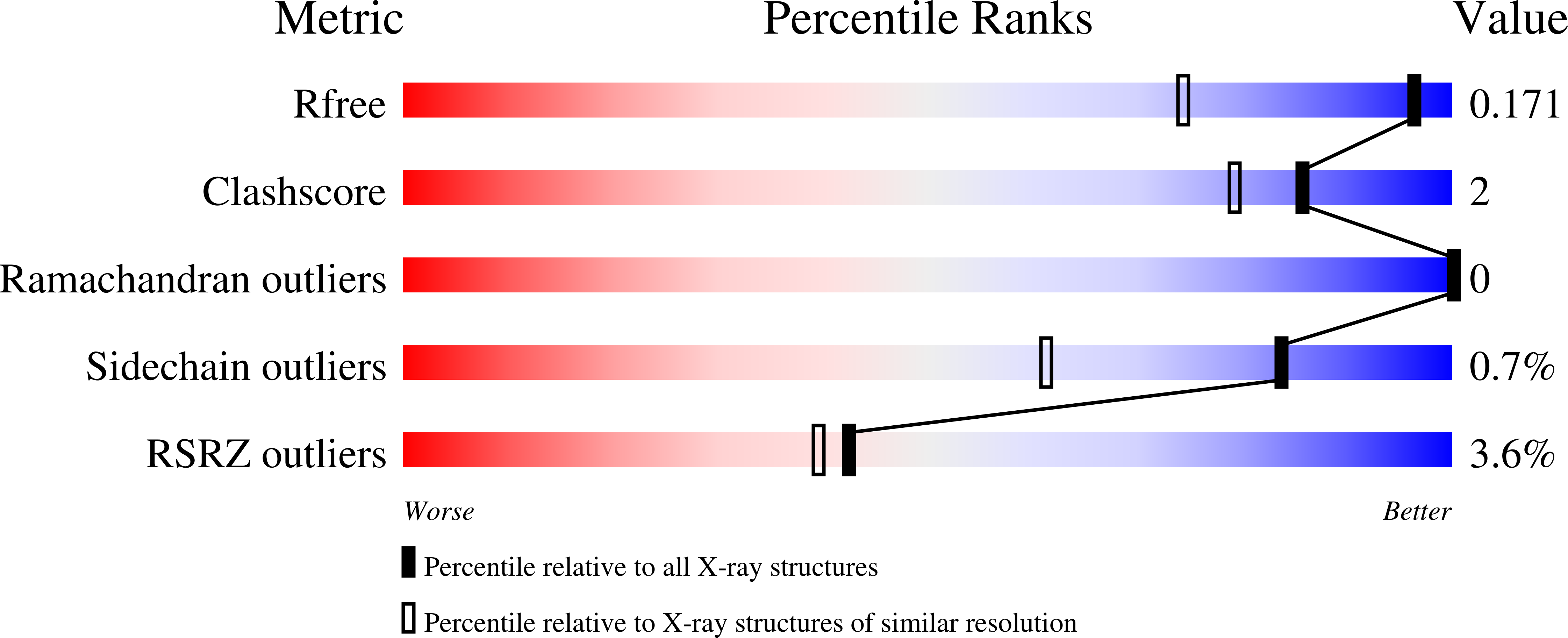
  • R-Value Free: 0.171 
  • R-Value Work: 0.149 
  • R-Value Observed: 0.150 

Starting Model: experimental
View more details

wwPDB Validation   3D Report Full Report


Ligand Structure Quality Assessment 


This is version 1.2 of the entry. See complete history


Literature

Identification of MRTX1133, a Noncovalent, Potent, and Selective KRAS G12D Inhibitor.

Wang, X.Allen, S.Blake, J.F.Bowcut, V.Briere, D.M.Calinisan, A.Dahlke, J.R.Fell, J.B.Fischer, J.P.Gunn, R.J.Hallin, J.Laguer, J.Lawson, J.D.Medwid, J.Newhouse, B.Nguyen, P.O'Leary, J.M.Olson, P.Pajk, S.Rahbaek, L.Rodriguez, M.Smith, C.R.Tang, T.P.Thomas, N.C.Vanderpool, D.Vigers, G.P.Christensen, J.G.Marx, M.A.

(2022) J Med Chem 65: 3123-3133

  • DOI: https://doi.org/10.1021/acs.jmedchem.1c01688
  • Primary Citation of Related Structures:  
    7RPZ, 7RT1, 7RT2, 7RT3, 7RT4, 7RT5

  • PubMed Abstract: 

    KRAS G12D , the most common oncogenic KRAS mutation, is a promising target for the treatment of solid tumors. However, when compared to KRAS G12C , selective inhibition of KRAS G12D presents a significant challenge due to the requirement of inhibitors to bind KRAS G12D with high enough affinity to obviate the need for covalent interactions with the mutant KRAS protein. Here, we report the discovery and characterization of the first noncovalent, potent, and selective KRAS G12D inhibitor, MRTX1133, which was discovered through an extensive structure-based activity improvement and shown to be efficacious in a KRAS G12D mutant xenograft mouse tumor model.


  • Organizational Affiliation

    Mirati Therapeutics, San Diego, California 92121, United States.


Macromolecules
Find similar proteins by:  (by identity cutoff)  |  3D Structure
Entity ID: 1
MoleculeChains Sequence LengthOrganismDetailsImage
Isoform 2B of GTPase KRas170Homo sapiensMutation(s): 1 
Gene Names: KRASKRAS2RASK2
EC: 3.6.5.2
UniProt & NIH Common Fund Data Resources
Find proteins for P01116 (Homo sapiens)
Explore P01116 
Go to UniProtKB:  P01116
PHAROS:  P01116
GTEx:  ENSG00000133703 
Entity Groups  
Sequence Clusters30% Identity50% Identity70% Identity90% Identity95% Identity100% Identity
UniProt GroupP01116
Sequence Annotations
Expand
  • Reference Sequence
Small Molecules
Ligands 3 Unique
IDChains Name / Formula / InChI Key2D Diagram3D Interactions
6IC (Subject of Investigation/LOI)
Query on 6IC

Download Ideal Coordinates CCD File 
C [auth A]4-(4-[(1R,5S)-3,8-diazabicyclo[3.2.1]octan-3-yl]-8-fluoro-2-{[(2R,4R,7aS)-2-fluorotetrahydro-1H-pyrrolizin-7a(5H)-yl]methoxy}pyrido[4,3-d]pyrimidin-7-yl)-5-ethynyl-6-fluoronaphthalen-2-ol
C33 H31 F3 N6 O2
SCLLZBIBSFTLIN-INOGPEIASA-N
GDP
Query on GDP

Download Ideal Coordinates CCD File 
B [auth A]GUANOSINE-5'-DIPHOSPHATE
C10 H15 N5 O11 P2
QGWNDRXFNXRZMB-UUOKFMHZSA-N
MG
Query on MG

Download Ideal Coordinates CCD File 
D [auth A]MAGNESIUM ION
Mg
JLVVSXFLKOJNIY-UHFFFAOYSA-N
Experimental Data & Validation

Experimental Data

  • Method: X-RAY DIFFRACTION
  • Resolution: 1.30 Å
  • R-Value Free: 0.171 
  • R-Value Work: 0.149 
  • R-Value Observed: 0.150 
  • Space Group: P 21 21 21
Unit Cell:
Length ( Å )Angle ( ˚ )
a = 39.868α = 90
b = 51.757β = 90
c = 89.611γ = 90
Software Package:
Software NamePurpose
PHENIXrefinement
PHASERphasing

Structure Validation

View Full Validation Report


Ligand Structure Quality Assessment 


Entry History & Funding Information

Deposition Data


Funding OrganizationLocationGrant Number
Not funded--

Revision History  (Full details and data files)

  • Version 1.0: 2021-12-22
    Type: Initial release
  • Version 1.1: 2022-03-09
    Changes: Database references
  • Version 1.2: 2023-10-18
    Changes: Data collection, Refinement description