7WR5

Crystal structure of OspC3-calmodulin-caspase-4 complex binding with 2'-aF-NAD+


Experimental Data Snapshot

  • Method: X-RAY DIFFRACTION
  • Resolution: 3.10 Å
  • R-Value Free: 0.272 
  • R-Value Work: 0.226 
  • R-Value Observed: 0.230 

wwPDB Validation   3D Report Full Report


Ligand Structure Quality Assessment 


This is version 1.3 of the entry. See complete history


Literature

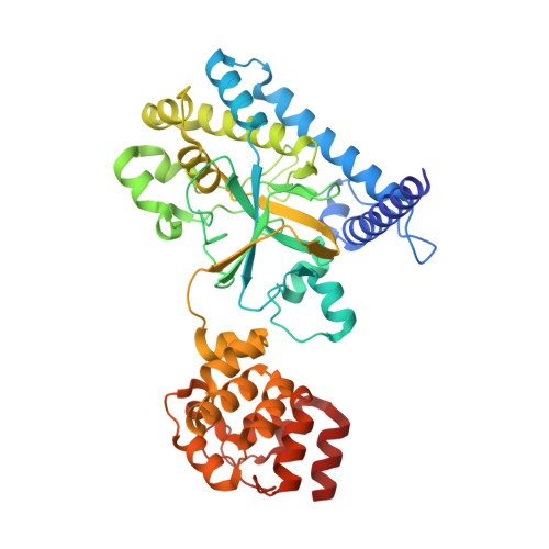
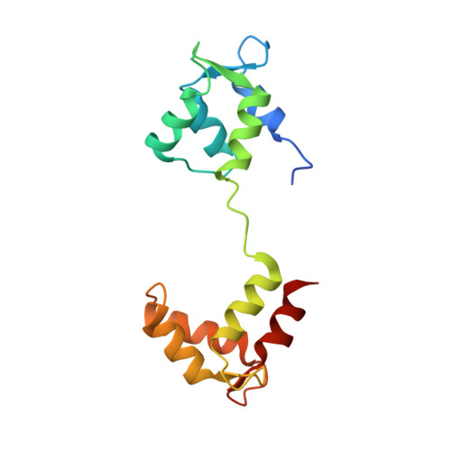
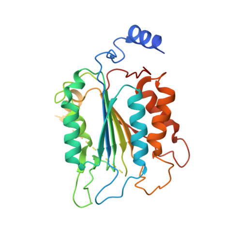
Structural mechanisms of calmodulin activation of Shigella effector OspC3 to ADP-riboxanate caspase-4/11 and block pyroptosis.

Hou, Y.Zeng, H.Li, Z.Feng, N.Meng, F.Xu, Y.Li, L.Shao, F.Ding, J.

(2023) Nat Struct Mol Biol 30: 261-272

  • DOI: https://doi.org/10.1038/s41594-022-00888-3
  • Primary Citation of Related Structures:  
    7WR0, 7WR1, 7WR2, 7WR3, 7WR4, 7WR5, 7WR6

  • PubMed Abstract: 

    The caspase-4/11-GSDMD pyroptosis axis recognizes cytosolic lipopolysaccharide for antibacterial defenses. Shigella flexneri employs an OspC3 effector to block pyroptosis by catalyzing NAD + -dependent arginine ADP-riboxanation of caspase-4/11. Here, we identify Ca 2+ -free calmodulin (CaM) that binds and stimulates OspC3 ADP-riboxanase activity. Crystal structures of OspC3-CaM and OspC3-caspase-4 binary complexes reveal unique CaM binding to an OspC3 N-terminal domain featuring an ADP-ribosyltransferase-like fold and specific recognition of caspase-4 by an OspC3 ankryin repeat domain, respectively. CaM-OspC3-caspase-4 ternary complex structures show that NAD + binding reorganizes the catalytic pocket, in which D231 and D177 activate the substrate arginine for initial ADP-ribosylation and ribosyl 2'-OH in the ADP-ribosylated arginine, respectively, for subsequent deamination. We also determine structures of unmodified and OspC3-ADP-riboxanated caspase-4. Mechanisms derived from this series of structures covering the entire process of OspC3 action are supported by biochemical analyses in vitro and functional validation in S. flexneri-infected mice.


  • Organizational Affiliation

    National Laboratory of Biomacromolecules, CAS Center for Excellence in Biomacromolecules, Institute of Biophysics, Chinese Academy of Sciences, Beijing, China.


Macromolecules
Find similar proteins by:  (by identity cutoff)  |  3D Structure
Entity ID: 1
MoleculeChains Sequence LengthOrganismDetailsImage
OspC3430Shigella flexneriMutation(s): 0 
Gene Names: ospC3
UniProt
Find proteins for A0A0H2US87 (Shigella flexneri)
Explore A0A0H2US87 
Go to UniProtKB:  A0A0H2US87
Entity Groups  
Sequence Clusters30% Identity50% Identity70% Identity90% Identity95% Identity100% Identity
UniProt GroupA0A0H2US87
Sequence Annotations
Expand
  • Reference Sequence
Find similar proteins by:  (by identity cutoff)  |  3D Structure
Entity ID: 2
MoleculeChains Sequence LengthOrganismDetailsImage
Calmodulin-1149Homo sapiensMutation(s): 0 
Gene Names: CALM1CALMCAMCAM1
UniProt & NIH Common Fund Data Resources
Find proteins for P0DP23 (Homo sapiens)
Explore P0DP23 
Go to UniProtKB:  P0DP23
PHAROS:  P0DP23
GTEx:  ENSG00000198668 
Entity Groups  
Sequence Clusters30% Identity50% Identity70% Identity90% Identity95% Identity100% Identity
UniProt GroupP0DP23
Sequence Annotations
Expand
  • Reference Sequence
Find similar proteins by:  (by identity cutoff)  |  3D Structure
Entity ID: 3
MoleculeChains Sequence LengthOrganismDetailsImage
Caspase-4280Homo sapiensMutation(s): 1 
Gene Names: CASP4
EC: 3.4.22.57
UniProt & NIH Common Fund Data Resources
Find proteins for P49662 (Homo sapiens)
Explore P49662 
Go to UniProtKB:  P49662
PHAROS:  P49662
GTEx:  ENSG00000196954 
Entity Groups  
Sequence Clusters30% Identity50% Identity70% Identity90% Identity95% Identity100% Identity
UniProt GroupP49662
Sequence Annotations
Expand
  • Reference Sequence
Small Molecules
Ligands 1 Unique
IDChains Name / Formula / InChI Key2D Diagram3D Interactions
5ZV (Subject of Investigation/LOI)
Query on 5ZV

Download Ideal Coordinates CCD File 
D [auth A][[(2~{R},3~{R},4~{S},5~{R})-5-(3-aminocarbonylpyridin-1-yl)-4-fluoranyl-3-oxidanyl-oxolan-2-yl]methoxy-oxidanyl-phosphoryl] [(2~{R},3~{S},4~{R},5~{R})-5-(6-aminopurin-9-yl)-3,4-bis(oxidanyl)oxolan-2-yl]methyl hydrogen phosphate
C21 H27 F N7 O13 P2
KKCPXEYWHWVQJS-IVHCLJLKSA-N
Experimental Data & Validation

Experimental Data

  • Method: X-RAY DIFFRACTION
  • Resolution: 3.10 Å
  • R-Value Free: 0.272 
  • R-Value Work: 0.226 
  • R-Value Observed: 0.230 
  • Space Group: P 2 21 21
Unit Cell:
Length ( Å )Angle ( ˚ )
a = 52.317α = 90
b = 120.074β = 90
c = 141.475γ = 90
Software Package:
Software NamePurpose
PHENIXrefinement
XDSdata reduction
XDSdata scaling
PHASERphasing

Structure Validation

View Full Validation Report


Ligand Structure Quality Assessment 


Entry History & Funding Information

Deposition Data


Funding OrganizationLocationGrant Number
Chinese Academy of SciencesChina--

Revision History  (Full details and data files)

  • Version 1.0: 2023-01-25
    Type: Initial release
  • Version 1.1: 2023-02-01
    Changes: Database references
  • Version 1.2: 2023-03-29
    Changes: Database references
  • Version 1.3: 2023-11-29
    Changes: Data collection, Refinement description