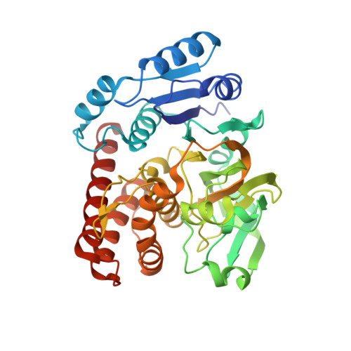
7XPS

Crystal structrue of MtdL:GDP:Mn


Experimental Data Snapshot

  • Method: X-RAY DIFFRACTION
  • Resolution: 2.10 Å
  • R-Value Free: 0.267 
  • R-Value Work: 0.224 
  • R-Value Observed: 0.226 

Starting Model: experimental
View more details

wwPDB Validation   3D Report Full Report


Ligand Structure Quality Assessment 


This is version 1.1 of the entry. See complete history


Literature

Structures of the NDP-pyranose mutase belonging to glycosyltransferase family 75 reveal residues important for Mn 2+ coordination and substrate binding.

Du, X.Chu, X.Liu, N.Jia, X.Peng, H.Xiao, Y.Liu, L.Yu, H.Li, F.He, C.

(2023) J Biol Chem 299: 102903-102903

  • DOI: https://doi.org/10.1016/j.jbc.2023.102903
  • Primary Citation of Related Structures:  
    7XPR, 7XPS, 7XPT, 7XPU, 7XPV, 8HL8

  • PubMed Abstract: 

    Members of glycosyltransferase family 75 (GT75) not only reversibly catalyze the autoglycosylation of a conserved arginine residue with specific NDP-sugars but also exhibit NDP-pyranose mutase activity that reversibly converts specific NDP-pyranose to NDP-furanose. The latter activity provides valuable NDP-furanosyl donors for glycosyltransferases and requires a divalent cation as a cofactor instead of FAD used by UDP-D-galactopyranose mutase. However, details of the mechanism for NDP-pyranose mutase activity are not clear. Here we report the first crystal structures of GT75 family NDP-pyranose mutases. The novel structures of GT75 member MtdL in complex with Mn 2+ and GDP, GDP-D-glucopyranose, GDP-L-fucopyranose, GDP-L-fucofuranose, respectively, combined with site-directed mutagenesis studies, reveal key residues involved in Mn 2+ coordination, substrate binding, and catalytic reactions. We also provide a possible catalytic mechanism for this unique type of NDP-pyranose mutase. Taken together, our results highlight key elements of an enzyme family important for furanose biosynthesis.


  • Organizational Affiliation

    Anhui Key Laboratory of Modern Biomanufacturing and School of Life Sciences, Anhui University, Hefei, Anhui, China.


Macromolecules
Find similar proteins by:  (by identity cutoff)  |  3D Structure
Entity ID: 1
MoleculeChains Sequence LengthOrganismDetailsImage
Transglycosylse
A, B
377Marinactinospora thermotoleransMutation(s): 0 
UniProt
Find proteins for G8HX37 (Marinactinospora thermotolerans)
Explore G8HX37 
Go to UniProtKB:  G8HX37
Entity Groups  
Sequence Clusters30% Identity50% Identity70% Identity90% Identity95% Identity100% Identity
UniProt GroupG8HX37
Sequence Annotations
Expand
  • Reference Sequence
Experimental Data & Validation

Experimental Data

  • Method: X-RAY DIFFRACTION
  • Resolution: 2.10 Å
  • R-Value Free: 0.267 
  • R-Value Work: 0.224 
  • R-Value Observed: 0.226 
  • Space Group: C 2 2 21
Unit Cell:
Length ( Å )Angle ( ˚ )
a = 98.694α = 90
b = 162.243β = 90
c = 116.496γ = 90
Software Package:
Software NamePurpose
PHENIXrefinement
Aimlessdata scaling
PDB_EXTRACTdata extraction
XDSdata reduction
PHASERphasing

Structure Validation

View Full Validation Report


Ligand Structure Quality Assessment 


Entry History & Funding Information

Deposition Data


Funding OrganizationLocationGrant Number
National Natural Science Foundation of China (NSFC)China--

Revision History  (Full details and data files)

  • Version 1.0: 2023-03-29
    Type: Initial release
  • Version 1.1: 2023-11-29
    Changes: Data collection, Refinement description