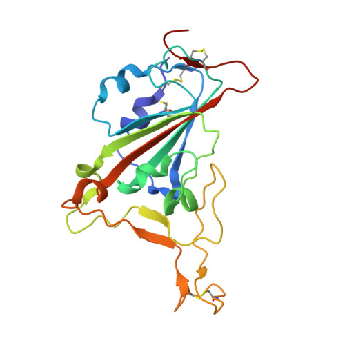
8EDF

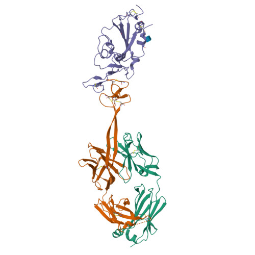
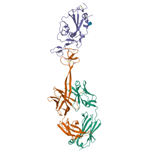
Bovine Fab SKD in complex with Sars COV-2 receptor binding domain


Experimental Data Snapshot

  • Method: X-RAY DIFFRACTION
  • Resolution: 3.40 Å
  • R-Value Free: 0.299 
  • R-Value Work: 0.276 
  • R-Value Observed: 0.277 

wwPDB Validation   3D Report Full Report


Ligand Structure Quality Assessment 


This is version 1.2 of the entry. See complete history


Literature

The smallest functional antibody fragment: Ultralong CDR H3 antibody knob regions potently neutralize SARS-CoV-2.

Huang, R.Warner Jenkins, G.Kim, Y.Stanfield, R.L.Singh, A.Martinez-Yamout, M.Kroon, G.J.Torres, J.L.Jackson, A.M.Kelley, A.Shaabani, N.Zeng, B.Bacica, M.Chen, W.Warner, C.Radoicic, J.Joh, J.Dinali Perera, K.Sang, H.Kim, T.Yao, J.Zhao, F.Sok, D.Burton, D.R.Allen, J.Harriman, W.Mwangi, W.Chung, D.Teijaro, J.R.Ward, A.B.Dyson, H.J.Wright, P.E.Wilson, I.A.Chang, K.O.McGregor, D.Smider, V.V.

(2023) Proc Natl Acad Sci U S A 120: e2303455120-e2303455120

  • DOI: https://doi.org/10.1073/pnas.2303455120
  • Primary Citation of Related Structures:  
    8ECQ, 8ECV, 8ECZ, 8ED1, 8EDF

  • PubMed Abstract: 

    Cows produce antibodies with a disulfide-bonded antigen-binding domain embedded within ultralong heavy chain third complementarity determining regions. This "knob" domain is analogous to natural cysteine-rich peptides such as knottins in that it is small and stable but can accommodate diverse loops and disulfide bonding patterns. We immunized cattle with SARS-CoV-2 spike and found ultralong CDR H3 antibodies that could neutralize several viral variants at picomolar IC 50 potencies in vitro and could protect from disease in vivo. The independent CDR H3 peptide knobs were expressed and maintained the properties of the parent antibodies. The knob interaction with SARS-CoV-2 spike was revealed by electron microscopy, X-ray crystallography, NMR spectroscopy, and mass spectrometry and established ultralong CDR H3-derived knobs as the smallest known recombinant independent antigen-binding fragment. Unlike other vertebrate antibody fragments, these knobs are not reliant on the immunoglobulin domain and have potential as a new class of therapeutics.


  • Organizational Affiliation

    Applied Biomedical Science Institute, San Diego, CA 92127.


Macromolecules
Find similar proteins by:  (by identity cutoff)  |  3D Structure
Entity ID: 1
MoleculeChains Sequence LengthOrganismDetailsImage
SKD Fab Light chainA [auth L]216Bos taurusMutation(s): 0 
UniProt & NIH Common Fund Data Resources
Find proteins for P0DOY2 (Homo sapiens)
Explore P0DOY2 
Go to UniProtKB:  P0DOY2
GTEx:  ENSG00000211677 
Entity Groups  
Sequence Clusters30% Identity50% Identity70% Identity90% Identity95% Identity100% Identity
UniProt GroupP0DOY2
Sequence Annotations
Expand
  • Reference Sequence
Find similar proteins by:  (by identity cutoff)  |  3D Structure
Entity ID: 2
MoleculeChains Sequence LengthOrganismDetailsImage
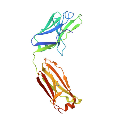
SKD Fab heavy chainB [auth H]272Bos taurusHomo sapiens
This entity is chimeric
Mutation(s): 0 
UniProt
Find proteins for P0DOX5 (Homo sapiens)
Explore P0DOX5 
Go to UniProtKB:  P0DOX5
Entity Groups  
Sequence Clusters30% Identity50% Identity70% Identity90% Identity95% Identity100% Identity
UniProt GroupP0DOX5
Sequence Annotations
Expand
  • Reference Sequence
Find similar proteins by:  (by identity cutoff)  |  3D Structure
Entity ID: 3
MoleculeChains Sequence LengthOrganismDetailsImage
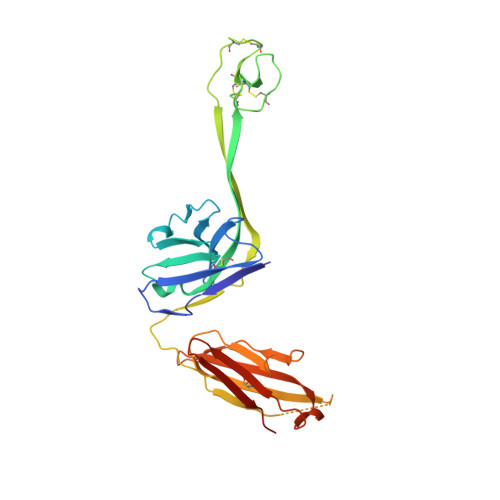
Spike protein S1C [auth A]200Severe acute respiratory syndrome coronavirus 2Mutation(s): 0 
Gene Names: S2
UniProt
Find proteins for P0DTC2 (Severe acute respiratory syndrome coronavirus 2)
Explore P0DTC2 
Go to UniProtKB:  P0DTC2
Entity Groups  
Sequence Clusters30% Identity50% Identity70% Identity90% Identity95% Identity100% Identity
UniProt GroupP0DTC2
Glycosylation
Glycosylation Sites: 1Go to GlyGen: P0DTC2-1
Sequence Annotations
Expand
  • Reference Sequence
Small Molecules
Ligands 1 Unique
IDChains Name / Formula / InChI Key2D Diagram3D Interactions
NAG
Query on NAG

Download Ideal Coordinates CCD File 
D [auth A]2-acetamido-2-deoxy-beta-D-glucopyranose
C8 H15 N O6
OVRNDRQMDRJTHS-FMDGEEDCSA-N
Experimental Data & Validation

Experimental Data

  • Method: X-RAY DIFFRACTION
  • Resolution: 3.40 Å
  • R-Value Free: 0.299 
  • R-Value Work: 0.276 
  • R-Value Observed: 0.277 
  • Space Group: P 41 21 2
Unit Cell:
Length ( Å )Angle ( ˚ )
a = 162.841α = 90
b = 162.841β = 90
c = 125.171γ = 90
Software Package:
Software NamePurpose
PHENIXrefinement
HKL-2000data reduction
HKL-2000data scaling
PHASERphasing

Structure Validation

View Full Validation Report


Ligand Structure Quality Assessment 


Entry History & Funding Information

Deposition Data


Funding OrganizationLocationGrant Number
Bill & Melinda Gates FoundationUnited StatesOPP1170236
Bill & Melinda Gates FoundationUnited StatesINV-004923

Revision History  (Full details and data files)

  • Version 1.0: 2023-09-20
    Type: Initial release
  • Version 1.1: 2023-10-04
    Changes: Database references
  • Version 1.2: 2024-10-16
    Changes: Structure summary