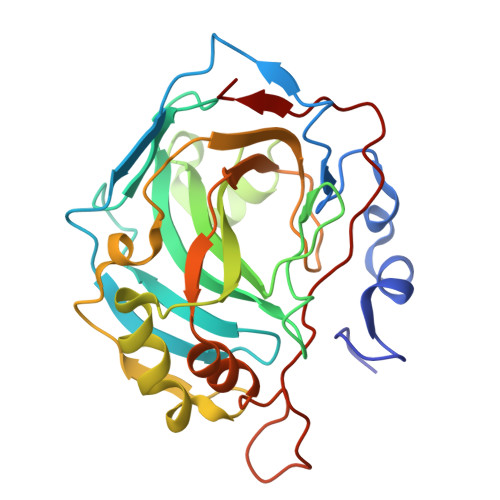
From X-ray crystallographic structure to intrinsic thermodynamics of protein-ligand binding using carbonic anhydrase isozymes as a model system.
Paketuryte-Latve, V., Smirnov, A., Manakova, E., Baranauskiene, L., Petrauskas, V., Zubriene, A., Matuliene, J., Dudutiene, V., Capkauskaite, E., Zaksauskas, A., Leitans, J., Grazulis, S., Tars, K., Matulis, D.(2024) IUCrJ 11: 556-569
- PubMed: 38856178
- DOI: https://doi.org/10.1107/S2052252524004627
- Primary Citation of Related Structures:
8RAR, 8RBP, 8RJ2 - PubMed Abstract:
Carbonic anhydrase (CA) was among the first proteins whose X-ray crystal structure was solved to atomic resolution. CA proteins have essentially the same fold and similar active centers that differ in only several amino acids. Primary sulfonamides are well defined, strong and specific binders of CA. However, minor variations in chemical structure can significantly alter their binding properties. Over 1000 sulfonamides have been designed, synthesized and evaluated to understand the correlations between the structure and thermodynamics of their binding to the human CA isozyme family. Compound binding was determined by several binding assays: fluorescence-based thermal shift assay, stopped-flow enzyme activity inhibition assay, isothermal titration calorimetry and competition assay for enzyme expressed on cancer cell surfaces. All assays have advantages and limitations but are necessary for deeper characterization of these protein-ligand interactions. Here, the concept and importance of intrinsic binding thermodynamics is emphasized and the role of structure-thermodynamics correlations for the novel inhibitors of CA IX is discussed - an isozyme that is overexpressed in solid hypoxic tumors, and thus these inhibitors may serve as anticancer drugs. The abundant structural and thermodynamic data are assembled into the Protein-Ligand Binding Database to understand general protein-ligand recognition principles that could be used in drug discovery.
Organizational Affiliation:
Department of Biothermodynamics and Drug Design, Institute of Biotechnology, Life Sciences Center, Vilnius University, Saulėtekio 7, 10257 Vilnius, Lithuania.


