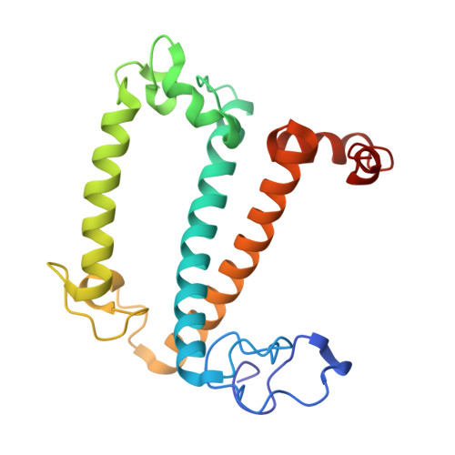
8Y15

Cryo-EM structure of Light-harvesting complex from Spinacia oleracea


Experimental Data Snapshot

  • Method: ELECTRON MICROSCOPY
  • Resolution: 2.17 Å
  • Aggregation State: PARTICLE 
  • Reconstruction Method: SINGLE PARTICLE 

Starting Model: experimental
View more details

wwPDB Validation   3D Report Full Report


This is version 1.1 of the entry. See complete history


Literature

Structure-based validation of recombinant light-harvesting complex II.

Seki, S.Miyata, T.Norioka, N.Tanaka, H.Kurisu, G.Namba, K.Fujii, R.

(2024) PNAS Nexus 3: pgae405-pgae405

  • DOI: https://doi.org/10.1093/pnasnexus/pgae405
  • Primary Citation of Related Structures:  
    8Y15, 8YEE

  • PubMed Abstract: 

    Light-harvesting complex II (LHCII) captures sunlight and dissipates excess energy to drive photosynthesis. To elucidate this mechanism, the individual optical properties of pigments in the LHCII protein must be identified. In vitro reconstitution with apoproteins synthesized by Escherichia coli and pigment-lipid mixtures from natural sources is an effective approach; however, the local environment surrounding each pigment within reconstituted LHCII (rLHCII) has only been indirectly estimated using spectroscopic and biochemical methods. Here, we used cryo-electron microscopy to determine the 3D structure of the rLHCII trimer and found that rLHCII exhibited a structure that was virtually identical to that of native LHCII, with a few exceptions: some C-terminal amino acids were not visible, likely due to aggregation of the His-tags; a carotenoid at the V1 site was not visible; and at site 614 showed mixed occupancy by both chlorophyll a and b molecules. Our observations confirmed the applicability of the in vitro reconstitution technique.


  • Organizational Affiliation

    Graduate School of Science, Osaka City University, 3-3-138 Sugimoto, Sumiyoshi-ku, Osaka 558-8585, Japan.


Macromolecules
Find similar proteins by:  (by identity cutoff)  |  3D Structure
Entity ID: 1
MoleculeChains Sequence LengthOrganismDetailsImage
Chlorophyll a-b binding protein, chloroplastic
A, B, C
216Spinacia oleraceaMutation(s): 0 
UniProt
Find proteins for P12333 (Spinacia oleracea)
Explore P12333 
Go to UniProtKB:  P12333
Entity Groups  
Sequence Clusters30% Identity50% Identity70% Identity90% Identity95% Identity100% Identity
UniProt GroupP12333
Sequence Annotations
Expand
  • Reference Sequence
Small Molecules
Ligands 6 Unique
IDChains Name / Formula / InChI Key2D Diagram3D Interactions
CHL (Subject of Investigation/LOI)
Query on CHL

Download Ideal Coordinates CCD File 
AA [auth B]
BA [auth B]
CA [auth B]
D [auth A]
DA [auth B]
AA [auth B],
BA [auth B],
CA [auth B],
D [auth A],
DA [auth B],
EA [auth B],
H [auth A],
I [auth A],
J [auth A],
K [auth A],
L [auth A],
QA [auth C],
UA [auth C],
VA [auth C],
W [auth B],
WA [auth C],
XA [auth C],
YA [auth C]
CHLOROPHYLL B
C55 H70 Mg N4 O6
MWVCRINOIIOUAU-UYSPMESUSA-M
CLA (Subject of Investigation/LOI)
Query on CLA

Download Ideal Coordinates CCD File 
AB [auth C]
BB [auth C]
CB [auth C]
DB [auth C]
E [auth A]
AB [auth C],
BB [auth C],
CB [auth C],
DB [auth C],
E [auth A],
F [auth A],
FA [auth B],
G [auth A],
GA [auth B],
HA [auth B],
IA [auth B],
JA [auth B],
M [auth A],
N [auth A],
O [auth A],
P [auth A],
Q [auth A],
RA [auth C],
SA [auth C],
TA [auth C],
X [auth B],
Y [auth B],
Z [auth B],
ZA [auth C]
CHLOROPHYLL A
C55 H72 Mg N4 O5
ATNHDLDRLWWWCB-AENOIHSZSA-M
LHG (Subject of Investigation/LOI)
Query on LHG

Download Ideal Coordinates CCD File 
HB [auth C],
NA [auth B],
U [auth A]
1,2-DIPALMITOYL-PHOSPHATIDYL-GLYCEROLE
C38 H75 O10 P
BIABMEZBCHDPBV-MPQUPPDSSA-N
XAT (Subject of Investigation/LOI)
Query on XAT

Download Ideal Coordinates CCD File 
OA [auth B],
PA [auth C],
V [auth A]
(3S,5R,6S,3'S,5'R,6'S)-5,6,5',6'-DIEPOXY-5,6,5',6'- TETRAHYDRO-BETA,BETA-CAROTENE-3,3'-DIOL
C40 H56 O4
SZCBXWMUOPQSOX-WVJDLNGLSA-N
NEX (Subject of Investigation/LOI)
Query on NEX

Download Ideal Coordinates CCD File 
GB [auth C],
MA [auth B],
T [auth A]
(1R,3R)-6-{(3E,5E,7E,9E,11E,13E,15E,17E)-18-[(1S,4R,6R)-4-HYDROXY-2,2,6-TRIMETHYL-7-OXABICYCLO[4.1.0]HEPT-1-YL]-3,7,12,16-TETRAMETHYLOCTADECA-1,3,5,7,9,11,13,15,17-NONAENYLIDENE}-1,5,5-TRIMETHYLCYCLOHEXANE-1,3-DIOL
C40 H56 O4
PGYAYSRVSAJXTE-OQASCVKESA-N
A1LXP (Subject of Investigation/LOI)
Query on A1LXP

Download Ideal Coordinates CCD File 
EB [auth C]
FB [auth C]
KA [auth B]
LA [auth B]
R [auth A]
EB [auth C],
FB [auth C],
KA [auth B],
LA [auth B],
R [auth A],
S [auth A]
Lutein
C40 H56 O2
KBPHJBAIARWVSC-RGZFRNHPSA-N
Experimental Data & Validation

Experimental Data

  • Method: ELECTRON MICROSCOPY
  • Resolution: 2.17 Å
  • Aggregation State: PARTICLE 
  • Reconstruction Method: SINGLE PARTICLE 
EM Software:
TaskSoftware PackageVersion
RECONSTRUCTIONcryoSPARC4.2.1
MODEL REFINEMENTCoot0.9.4.1
MODEL REFINEMENTPHENIX1.19.2

Structure Validation

View Full Validation Report


Entry History & Funding Information

Deposition Data


Funding OrganizationLocationGrant Number
Japan Society for the Promotion of Science (JSPS)JapanK23KJ1834
Japan Agency for Medical Research and Development (AMED)JapanJP23ama121003
Japan Science and TechnologyJapanJPMJFS2138
Japan Science and TechnologyJapanJPMJCR20E1
Japan Society for the Promotion of Science (JSPS)Japan23H04958

Revision History  (Full details and data files)

  • Version 1.0: 2024-11-20
    Type: Initial release
  • Version 1.1: 2024-12-11
    Changes: Advisory, Data collection, Derived calculations, Structure summary