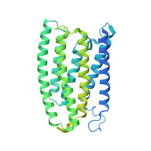
8YEK

Cryo-EM structure of the channelrhodopsin GtCCR2


Experimental Data Snapshot

  • Method: ELECTRON MICROSCOPY
  • Resolution: 2.73 Å
  • Aggregation State: PARTICLE 
  • Reconstruction Method: SINGLE PARTICLE 

Starting Model: in silico
View more details

wwPDB Validation   3D Report Full Report


This is version 1.3 of the entry. See complete history


Literature

The high-light-sensitivity mechanism and optogenetic properties of the bacteriorhodopsin-like channelrhodopsin GtCCR4.

Tanaka, T.Hososhima, S.Yamashita, Y.Sugimoto, T.Nakamura, T.Shigemura, S.Iida, W.Sano, F.K.Oda, K.Uchihashi, T.Katayama, K.Furutani, Y.Tsunoda, S.P.Shihoya, W.Kandori, H.Nureki, O.

(2024) Mol Cell 84: 3530-3544.e6

  • DOI: https://doi.org/10.1016/j.molcel.2024.08.016
  • Primary Citation of Related Structures:  
    8YEJ, 8YEK, 8YEL

  • PubMed Abstract: 

    Channelrhodopsins are microbial light-gated ion channels that can control the firing of neurons in response to light. Among several cation channelrhodopsins identified in Guillardia theta (GtCCRs), GtCCR4 has higher light sensitivity than typical channelrhodopsins. Furthermore, GtCCR4 shows superior properties as an optogenetic tool, such as minimal desensitization. Our structural analyses of GtCCR2 and GtCCR4 revealed that GtCCR4 has an outwardly bent transmembrane helix, resembling the conformation of activated G-protein-coupled receptors. Spectroscopic and electrophysiological comparisons suggested that this helix bend in GtCCR4 omits channel recovery time and contributes to high light sensitivity. An electrophysiological comparison of GtCCR4 and the well-characterized optogenetic tool ChRmine demonstrated that GtCCR4 has superior current continuity and action-potential spike generation with less invasiveness in neurons. We also identified highly active mutants of GtCCR4. These results shed light on the diverse structures and dynamics of microbial rhodopsins and demonstrate the strong optogenetic potential of GtCCR4.


  • Organizational Affiliation

    Department of Biological Sciences, Graduate School of Science, The University of Tokyo, Bunkyo, Tokyo 113-0033, Japan.


Macromolecules
Find similar proteins by:  (by identity cutoff)  |  3D Structure
Entity ID: 1
MoleculeChains Sequence LengthOrganismDetailsImage
GtCCR2464Guillardia theta CCMP2712Mutation(s): 0 
Gene Names: GUITHDRAFT_99928
Membrane Entity: Yes 
UniProt
Find proteins for L1K1K8 (Guillardia theta (strain CCMP2712))
Explore L1K1K8 
Go to UniProtKB:  L1K1K8
Entity Groups  
Sequence Clusters30% Identity50% Identity70% Identity90% Identity95% Identity100% Identity
UniProt GroupL1K1K8
Sequence Annotations
Expand
  • Reference Sequence
Experimental Data & Validation

Experimental Data

  • Method: ELECTRON MICROSCOPY
  • Resolution: 2.73 Å
  • Aggregation State: PARTICLE 
  • Reconstruction Method: SINGLE PARTICLE 
EM Software:
TaskSoftware PackageVersion
MODEL REFINEMENTPHENIX
MODEL REFINEMENTServalcat
RECONSTRUCTIONRELION3.1

Structure Validation

View Full Validation Report


Entry History & Funding Information

Deposition Data


Funding OrganizationLocationGrant Number
Japan Society for the Promotion of Science (JSPS)Japan22KJ0577
Japan Society for the Promotion of Science (JSPS)Japan19H05777
Japan Society for the Promotion of Science (JSPS)Japan22K19371
Japan Society for the Promotion of Science (JSPS)Japan22H02751
Japan Society for the Promotion of Science (JSPS)Japan21H05037
Japan Agency for Medical Research and Development (AMED)JapanJP233fa627001
Japan Agency for Medical Research and Development (AMED)JapanJP23ama121002
Japan Agency for Medical Research and Development (AMED)JapanJP23ama121012

Revision History  (Full details and data files)

  • Version 1.0: 2024-09-04
    Type: Initial release
  • Version 1.1: 2024-09-18
    Changes: Data collection, Database references
  • Version 1.2: 2024-10-02
    Changes: Data collection, Database references
  • Version 1.3: 2024-11-06
    Changes: Data collection, Structure summary