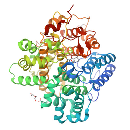
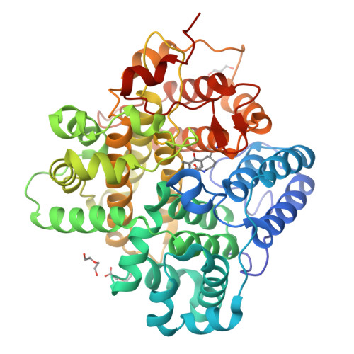
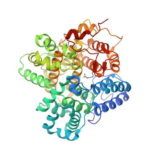
RCSB PDB - 8Z4S: The crystal structure of a Hydroquinone Dioxygenase PaD with nonnatural substrate S6

 8Z4S

The crystal structure of a Hydroquinone Dioxygenase PaD with nonnatural substrate S6


Experimental Data Snapshot

  • Method: X-RAY DIFFRACTION
  • Resolution: 1.93 Å
  • R-Value Free: 0.250 
  • R-Value Work: 0.209 
  • R-Value Observed: 0.211 

Starting Model: in silico
View more details

wwPDB Validation   3D Report Full Report


Ligand Structure Quality Assessment 

Created with Raphaël 2.3.0Worse 01 BetterLigand structure goodness of fit to experimental dataBest fitted A1D7ZClick on this verticalbar to view details

This is version 1.0 of the entry. See complete history


Literature

Substrate specificity of a branch of aromatic dioxygenases determined by three distinct motifs.

Cui, C.Yang, L.J.Liu, Z.W.Shu, X.Zhang, W.W.Gao, Y.Wang, Y.X.Wang, T.Chen, C.C.Guo, R.T.Gao, S.S.

(2024) Nat Commun 15: 7682-7682

  • DOI: https://doi.org/10.1038/s41467-024-52101-2
  • Primary Citation of Related Structures:  
    8Z4Q, 8Z4R, 8Z4S

  • PubMed Abstract: 

    The inversion of substrate size specificity is an evolutionary roadblock for proteins. The Duf4243 dioxygenases GedK and BTG13 are known to catalyze the aromatic cleavage of bulky tricyclic hydroquinone. In this study, we discover a Duf4243 dioxygenase PaD that favors small monocyclic hydroquinones from the penicillic-acid biosynthetic pathway. Sequence alignments between PaD and GedK and BTG13 suggest PaD has three additional motifs, namely motifs 1-3, distributed at different positions in the protein sequence. X-ray crystal structures of PaD with the substrate at high resolution show motifs 1-3 determine three loops (loops 1-3). Most intriguing, loops 1-3 stack together at the top of the pocket, creating a lid-like tertiary structure with a narrow channel and a clearly constricted opening. This drastically changes the substrate specificity by determining the entry and binding of much smaller substrates. Further genome mining suggests Duf4243 dioxygenases with motifs 1-3 belong to an evolutionary branch that is extensively involved in the biosynthesis of natural products and has the ability to degrade diverse monocyclic hydroquinone pollutants. This study showcases how natural enzymes alter the substrate specificity fundamentally by incorporating new small motifs, with a fixed overall scaffold-architecture. It will also offer a theoretical foundation for the engineering of substrate specificity in enzymes and act as a guide for the identification of aromatic dioxygenases with distinct substrate specificities.


  • Organizational Affiliation

    Tianjin Institute of Industrial Biotechnology, Chinese Academy of Sciences, Tianjin, China.


Macromolecules
Find similar proteins by:  (by identity cutoff)  |  3D Structure
Entity ID: 1
MoleculeChains Sequence LengthOrganismDetailsImage
Hydroquinone Dioxygenase PaD473Aspergillus westerdijkiaeMutation(s): 0 
Entity Groups  
Sequence Clusters30% Identity50% Identity70% Identity90% Identity95% Identity100% Identity
Sequence Annotations
Expand
  • Reference Sequence
Small Molecules
Ligands 6 Unique
IDChains Name / Formula / InChI Key2D Diagram3D Interactions
A1D7Z (Subject of Investigation/LOI)
Query on A1D7Z

Download Ideal Coordinates CCD File 
B [auth A]2,3,5-trimethylbenzene-1,4-diol
C9 H12 O2
AUFZRCJENRSRLY-UHFFFAOYSA-N
PEG
Query on PEG

Download Ideal Coordinates CCD File 
E [auth A],
F [auth A]
DI(HYDROXYETHYL)ETHER
C4 H10 O3
MTHSVFCYNBDYFN-UHFFFAOYSA-N
GOL
Query on GOL

Download Ideal Coordinates CCD File 
I [auth A]GLYCEROL
C3 H8 O3
PEDCQBHIVMGVHV-UHFFFAOYSA-N
ACT
Query on ACT

Download Ideal Coordinates CCD File 
D [auth A]ACETATE ION
C2 H3 O2
QTBSBXVTEAMEQO-UHFFFAOYSA-M
FE (Subject of Investigation/LOI)
Query on FE

Download Ideal Coordinates CCD File 
C [auth A]FE (III) ION
Fe
VTLYFUHAOXGGBS-UHFFFAOYSA-N
FMT
Query on FMT

Download Ideal Coordinates CCD File 
G [auth A],
H [auth A]
FORMIC ACID
C H2 O2
BDAGIHXWWSANSR-UHFFFAOYSA-N
Modified Residues  1 Unique
IDChains TypeFormula2D DiagramParent
KCX
Query on KCX
A
L-PEPTIDE LINKINGC7 H14 N2 O4LYS
Experimental Data & Validation

Experimental Data

  • Method: X-RAY DIFFRACTION
  • Resolution: 1.93 Å
  • R-Value Free: 0.250 
  • R-Value Work: 0.209 
  • R-Value Observed: 0.211 
  • Space Group: P 62
Unit Cell:
Length ( Å )Angle ( ˚ )
a = 119.831α = 90
b = 119.831β = 90
c = 92.889γ = 120
Software Package:
Software NamePurpose
REFMACrefinement
SAINTdata scaling
SAINTdata reduction
PHASERphasing

Structure Validation

View Full Validation Report


Ligand Structure Quality Assessment 

Created with Raphaël 2.3.0Worse 01 BetterLigand structure goodness of fit to experimental dataBest fitted A1D7ZClick on this verticalbar to view details

Entry History & Funding Information

Deposition Data


Funding OrganizationLocationGrant Number
National Natural Science Foundation of China (NSFC)China--

Revision History  (Full details and data files)

  • Version 1.0: 2024-09-11
    Type: Initial release