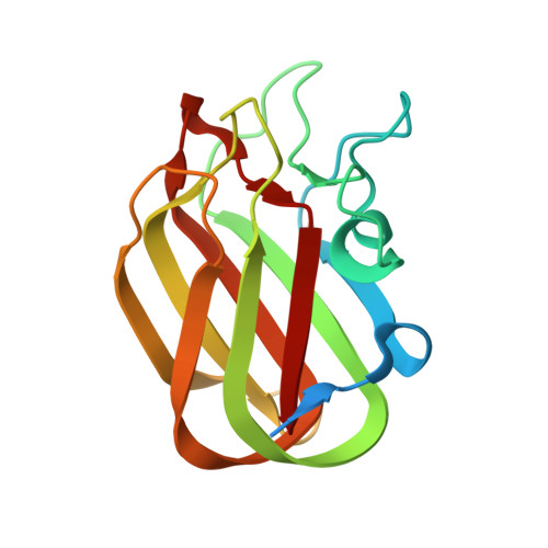
4A44

CpGH89CBM32-5, from Clostridium perfringens, in complex with the Tn Antigen


Experimental Data Snapshot

  • Method: X-RAY DIFFRACTION
  • Resolution: 1.70 Å
  • R-Value Free: 0.222 
  • R-Value Work: 0.188 
  • R-Value Observed: 0.189 

wwPDB Validation   3D Report Full Report


Ligand Structure Quality Assessment 


This is version 3.2 of the entry. See complete history


Literature

Carbohydrate Recognition by an Architecturally Complex Alpha-N-Acetylglucosaminidase from Clostridium Perfringens.

Ficko-Blean, E.Stuart, C.P.Suits, M.D.Cid, M.Tessier, M.Woods, R.J.Boraston, A.B.

(2012) PLoS One 7: 33524

  • DOI: https://doi.org/10.1371/journal.pone.0033524
  • Primary Citation of Related Structures:  
    4A3Z, 4A41, 4A42, 4A44, 4A45, 4A6O, 4AAX

  • PubMed Abstract: 

    CpGH89 is a large multimodular enzyme produced by the human and animal pathogen Clostridium perfringens. The catalytic activity of this exo-α-D-N-acetylglucosaminidase is directed towards a rare carbohydrate motif, N-acetyl-β-D-glucosamine-α-1,4-D-galactose, which is displayed on the class III mucins deep within the gastric mucosa. In addition to the family 89 glycoside hydrolase catalytic module this enzyme has six modules that share sequence similarity to the family 32 carbohydrate-binding modules (CBM32s), suggesting the enzyme has considerable capacity to adhere to carbohydrates. Here we suggest that two of the modules, CBM32-1 and CBM32-6, are not functional as carbohydrate-binding modules (CBMs) and demonstrate that three of the CBMs, CBM32-3, CBM32-4, and CBM32-5, are indeed capable of binding carbohydrates. CBM32-3 and CBM32-4 have a novel binding specificity for N-acetyl-β-D-glucosamine-α-1,4-D-galactose, which thus complements the specificity of the catalytic module. The X-ray crystal structure of CBM32-4 in complex with this disaccharide reveals a mode of recognition that is based primarily on accommodation of the unique bent shape of this sugar. In contrast, as revealed by a series of X-ray crystal structures and quantitative binding studies, CBM32-5 displays the structural and functional features of galactose binding that is commonly associated with CBM family 32. The functional CBM32s that CpGH89 contains suggest the possibility for multivalent binding events and the partitioning of this enzyme to highly specific regions within the gastrointestinal tract.


  • Organizational Affiliation

    Biochemistry and Microbiology, University of Victoria, Victoria, British Columbia, Canada.


Macromolecules
Find similar proteins by:  (by identity cutoff)  |  3D Structure
Entity ID: 1
MoleculeChains Sequence LengthOrganismDetailsImage
Alpha-N-acetylglucosaminidase161Clostridium perfringensMutation(s): 0 
Gene Names: CYK96_07800
UniProt
Find proteins for Q8XM24 (Clostridium perfringens (strain 13 / Type A))
Explore Q8XM24 
Go to UniProtKB:  Q8XM24
Entity Groups  
Sequence Clusters30% Identity50% Identity70% Identity90% Identity95% Identity100% Identity
UniProt GroupQ8XM24
Sequence Annotations
Expand
  • Reference Sequence
Small Molecules
Ligands 5 Unique
IDChains Name / Formula / InChI Key2D Diagram3D Interactions
A2G
Query on A2G

Download Ideal Coordinates CCD File 
H [auth A]2-acetamido-2-deoxy-alpha-D-galactopyranose
C8 H15 N O6
OVRNDRQMDRJTHS-CBQIKETKSA-N
CIT
Query on CIT

Download Ideal Coordinates CCD File 
F [auth A]CITRIC ACID
C6 H8 O7
KRKNYBCHXYNGOX-UHFFFAOYSA-N
SER
Query on SER

Download Ideal Coordinates CCD File 
G [auth A]SERINE
C3 H7 N O3
MTCFGRXMJLQNBG-REOHCLBHSA-N
CA
Query on CA

Download Ideal Coordinates CCD File 
B [auth A]CALCIUM ION
Ca
BHPQYMZQTOCNFJ-UHFFFAOYSA-N
NA
Query on NA

Download Ideal Coordinates CCD File 
C [auth A],
D [auth A],
E [auth A]
SODIUM ION
Na
FKNQFGJONOIPTF-UHFFFAOYSA-N
Experimental Data & Validation

Experimental Data

  • Method: X-RAY DIFFRACTION
  • Resolution: 1.70 Å
  • R-Value Free: 0.222 
  • R-Value Work: 0.188 
  • R-Value Observed: 0.189 
  • Space Group: P 21 21 21
Unit Cell:
Length ( Å )Angle ( ˚ )
a = 31.8α = 90
b = 59.26β = 90
c = 67.43γ = 90
Software Package:
Software NamePurpose
REFMACrefinement
d*TREKdata reduction
d*TREKdata scaling

Structure Validation

View Full Validation Report


Ligand Structure Quality Assessment 


Entry History 

Deposition Data

Revision History  (Full details and data files)

  • Version 1.0: 2012-04-04
    Type: Initial release
  • Version 1.1: 2012-04-18
    Changes: Other
  • Version 2.0: 2020-07-01
    Changes: Advisory, Atomic model, Data collection, Database references, Derived calculations, Non-polymer description, Other, Source and taxonomy, Structure summary
  • Version 3.0: 2020-07-29
    Type: Remediation
    Reason: Carbohydrate remediation
    Changes: Atomic model, Data collection, Derived calculations, Structure summary
  • Version 3.1: 2022-12-07
    Changes: Database references, Derived calculations, Structure summary
  • Version 3.2: 2024-06-19
    Changes: Data collection