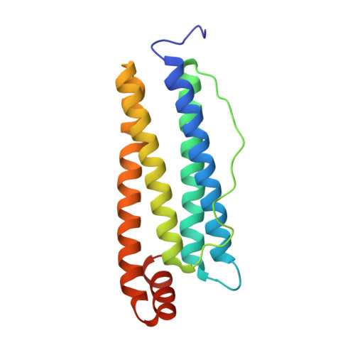
6HJU

The X-ray structure of the horse spleen ferritin nanocage containing Pt, obtained upon encapsulation of a Pt(II) terpyridine compound within the protein cage


Experimental Data Snapshot

  • Method: X-RAY DIFFRACTION
  • Resolution: 1.58 Å
  • R-Value Free: 0.182 
  • R-Value Work: 0.132 
  • R-Value Observed: 0.134 

wwPDB Validation   3D Report Full Report


This is version 1.2 of the entry. See complete history


Literature

Preparation, structure, cytotoxicity and mechanism of action of ferritin-Pt(II) terpyridine compound nanocomposites.

Ferraro, G.Pica, A.Petruk, G.Pane, F.Amoresano, A.Cilibrizzi, A.Vilar, R.Monti, D.M.Merlino, A.

(2018) Nanomedicine (Lond) 13: 2995-3007

  • DOI: https://doi.org/10.2217/nnm-2018-0259
  • Primary Citation of Related Structures:  
    6HJT, 6HJU

  • PubMed Abstract: 

    A Pt(II)-terpyridine compound, bearing two piperidine substituents at positions 2 and 2' of the terpyridine ligand (1), is highly cytotoxic and shows a mechanism of action distinct from cisplatin. 1 has been incorporated within the ferritin nanocage (AFt). Spectroscopic and crystallographic data of the Pt(II)-AFt nanocomposite have been collected and in vitro anticancer activity has been explored using cancer cells. Pt(II)-containing fragments bind His49, His114 and His132. Pt(II)-AFt nanocomposite is less cytotoxic than 1, but it is more toxic than cisplatin at high concentrations. The Pt(II)-AFt nanocomposite triggers necrosis in cancer cells, as free 1 does. Pt(II)-AFt nanocomposites are promising vehicles to deliver Pt-based drugs to cancer cells.


  • Organizational Affiliation

    Department of Chemical Sciences, University of Naples Federico II, Napoli, Italy.


Macromolecules
Find similar proteins by:  (by identity cutoff)  |  3D Structure
Entity ID: 1
MoleculeChains Sequence LengthOrganismDetailsImage
Ferritin light chain174Equus caballusMutation(s): 0 
UniProt
Find proteins for P02791 (Equus caballus)
Go to UniProtKB:  P02791
Sequence Annotations
Expand
  • Reference Sequence
Small Molecules
Ligands 6 Unique
IDChains Name / Formula / InChI Key2D Diagram3D Interactions
PT
Query on PT

Download Ideal Coordinates CCD File 
K [auth A],
L [auth A],
M [auth A],
N [auth A],
O [auth A]
PLATINUM (II) ION
Pt
HRGDZIGMBDGFTC-UHFFFAOYSA-N
CD
Query on CD

Download Ideal Coordinates CCD File 
B [auth A]
C [auth A]
D [auth A]
E [auth A]
F [auth A]
B [auth A],
C [auth A],
D [auth A],
E [auth A],
F [auth A],
G [auth A],
H [auth A]
CADMIUM ION
Cd
WLZRMCYVCSSEQC-UHFFFAOYSA-N
SO4
Query on SO4

Download Ideal Coordinates CCD File 
P [auth A]SULFATE ION
O4 S
QAOWNCQODCNURD-UHFFFAOYSA-L
GOL
Query on GOL

Download Ideal Coordinates CCD File 
Q [auth A]GLYCEROL
C3 H8 O3
PEDCQBHIVMGVHV-UHFFFAOYSA-N
DMS
Query on DMS

Download Ideal Coordinates CCD File 
R [auth A]DIMETHYL SULFOXIDE
C2 H6 O S
IAZDPXIOMUYVGZ-UHFFFAOYSA-N
CL
Query on CL

Download Ideal Coordinates CCD File 
I [auth A],
J [auth A],
S [auth A]
CHLORIDE ION
Cl
VEXZGXHMUGYJMC-UHFFFAOYSA-M
Experimental Data & Validation

Experimental Data

  • Method: X-RAY DIFFRACTION
  • Resolution: 1.58 Å
  • R-Value Free: 0.182 
  • R-Value Work: 0.132 
  • R-Value Observed: 0.134 
  • Space Group: F 4 3 2
Unit Cell:
Length ( Å )Angle ( ˚ )
a = 180.53α = 90
b = 180.53β = 90
c = 180.53γ = 90
Software Package:
Software NamePurpose
REFMACrefinement
XDSdata reduction
XSCALEdata scaling
BUSTERphasing

Structure Validation

View Full Validation Report


Entry History 

Deposition Data

Revision History  (Full details and data files)

  • Version 1.0: 2018-12-19
    Type: Initial release
  • Version 1.1: 2019-03-06
    Changes: Data collection, Database references
  • Version 1.2: 2024-05-15
    Changes: Data collection, Database references, Derived calculations